Advertisements
Advertisements
प्रश्न
Draw a diagram of the human eye as seen in a vertical section and label the parts which suit the following descriptions relating to the:
(i) photosensitive layer of the eye.
(ii) structure which is responsible for holding the eye lens in its position.
(iii) structure which maintains the shape of the eyeball and the area of no vision.
(iv) anterior chamber seen in front of the eye lens.
(v) outermost transparent layer seen in front of the eyeball.
उत्तर

APPEARS IN
संबंधित प्रश्न
Write the function of retina in human eye.
The size of the pupil becomes ______ when you see in dim light.
What kind of lens is present in the human eye?
What is the name of:
the light-sensitive layer in the eye?
Draw a simple diagram of the human eye and label clearly the cornea, iris, pupil, ciliary muscles, eye-lens, retina, optic nerve and blind spot.
What shape are your eye-lenses:
when you look at your hand?
Nocturnal animals (animals which sleep during the day and come out at night) tend to have wide pupils and lot of rods in their retinas. Suggest reasons for this.
Which of the following have a wider field of view?
(a) Animals having two eyes on the opposite sides of their head.
(b) Animals having two eyes at the front of their head.
The animals of prey have:
(a) two eyes at the front
(b) two eyes at the back
(c) two eyes on the sides
(d) one eye at the front and one on the side
The figure below compares a part of our eye with a part of a photographic camera.
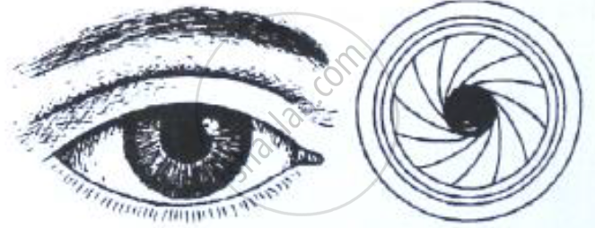
Name the corresponding parts of the eye the camera shown here that are comparable in function.
Correct and rewrite the statements by changing the biological term that is underlined for the statement:
The part of the eye which can be donated from a clinically dead person is the Retina.
The figure below compares a part of our eye with a part of a photographic camera.
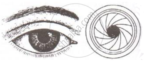
Explain the mode of working and the functions of the parts of the eye mentioned above.
Differentiate between:
Retina and Choroid.
Draw a labeled diagram of the front view of human eye.
State the Function:
Aqueous humour
Assertion: Blind spot is a small area of the retina which is insensitive to light where the optic nerve leaves the eye.
Reason: There are no rods or cones present at the junction of the optic nerve and retina in the eye.
Given below is a diagram depicting a defect of the human eye. Answer the questions that follow:
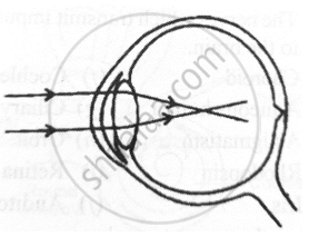
- Give the scientific term for the defect.
- Mention one possible reason for the defect.
- What type of lens can be used to correct the defect?
What kind of lens is there in our eyes? Where does it form the image of an object?
Write down the names of parts of the eye in the blank spaces shown in the figure.

Match the following:
|
Column - I |
Column - II | ||
| 1 | Retina | a | Path way of light |
| 2 | Pupil | b |
far point comes closer |
| 3 | Ciliary muscles | c |
near point moves away |
| 4 | Myopia | d | Screen of the eye |
| 5 | Hypermetropia | e | Power of accommodation |
