Advertisements
Advertisements
प्रश्न
Given alongside is a diagram of human heart showing its internal structures. Label the parts marked 1 to 6, and answer the following questions.

- Which type of blood is carried by the blood vessel marked 2?
- Name the main artery which takes the blood from heart to different parts of the body?
- Which chamber of the heart receives deoxygenated blood from the body?
उत्तर
- Left pulmonary artery
- Superior vena cava
- Left pulmonary vein
- Pericardium
- Left Ventricle
- Right Ventricle
- The superior vena cava carries deoxygenated blood from the upper part of the body (head, neck, and arms) back to the right atrium of the heart.
- The main artery that takes oxygenated blood from the heart to different parts of the body is the aorta
- The right atrium of the heart receives deoxygenated blood from the body.
APPEARS IN
संबंधित प्रश्न
Explain with tile help of a chart, explain the compatibility of human blood groups.
the Biological/technical term for Blood vessels carrying blood to the left atrium.
Give appropriate biological or technical terms for the following :
The complex consisting of a DNA strand and a core of histones
Name the following:
The smallest common blood vessels formed by the union of capillaries.
Name the following:
The fluid found between the membranes of the heart.
What does the term “double circulation” mean?
What is haemoglobin? State the consequences of deficiency of haemoglobin in our bodies.
Spleen is referred to as ______.
Name these:
Three circulating fluids in human body.
Given diagram is a schematic representation of the circulatory system in humans. Study the same and answer the questions that follow:
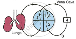 |
- Label the parts 1 and 4 indicated in the diagram.
- Which of the above mentioned number is the thickest artery? Also write its name.
- Mention the number and chamber of the heart which has the thickest muscular wall.
- Which of the above numbers/structures has the maximum number of blood capillaries?
- Draw neat and labelled diagrams of the transverse section of vena cava and the part numbered as 3. Make sure to show the structural differences between these two in the diagram.
