Advertisements
Advertisements
प्रश्न
Draw a neat and labelled diagram of the human ear. With the help of this diagram, explain the construction and working of the human ear.
उत्तर
Construction of the human ear is as follows:
The ear consists of three compartments: outer ear, middle ear, and inner ear. The outer ear consists of broad part called pinna and about 2–3 cm long tube called ear canal. At the end of ear canal is a thin circular elastic membrane called tympanum or eardrum. The middle ear contains three small delicate bones called hammer, anvil, and stirrup. These bones are linked to one another. The one end of the hammer touches the eardrum and the free end of the stirrup is held against the membrane over the oval window of the inner ear. The lower part of middle ear has Eustachian tube going to the throat. The inner ear has a coiled structure called cochlea. The cochlea is filled with liquid containing sound-sensitive nerve cells. The cochlea is connected to the auditory nerve that goes to the brain.
Working of the ear can be explained as follows:
The sound waves are collected by the pinna. These sound waves pass through the ear canal and fall on the eardrum. Sound waves consist of compressions and rarefactions. When the compression strikes the eardrum, the pressure on the outer surface of the eardrum increases and pushes the eardrum inward. Whereas when rarefaction strikes the eardrum, the pressure on the outer surface of eardrum decreases and pushes the eardrum outward. Thus, when sound falls on the eardrum, it vibrates back and forth rapidly. These vibrations are passed onto the three bones in the middle ear and finally to the liquid in the cochlea. Due to this phenomenon the liquid starts to vibrate, setting up electrical impulses in the nerve cells present in it. These impulses are carried to the brain by auditory nerves. The brain interprets the impulses and thus we feel the hearing sensation.
APPEARS IN
संबंधित प्रश्न
Draw labelled diagrams of the following: Ear
Eye : Optic nerve : : Ear : ___________
Differentiate between members of the following pair with reference to what is asked in bracket.
Dynamic balance and static balance (Definition)
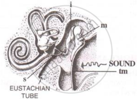
The figure below is the sectional view of a part of the skull showing a sense organ:

What are the parts labeled 'm', 'i' and 's'? What do these parts constitute collectively?
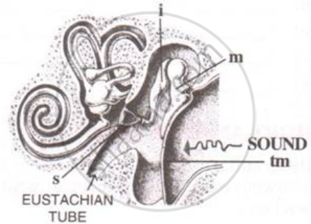
The figure below is the sectional view of a part of the skull showing a sense organ:

Name the part labeled 'tm'. What is its function?
Given below is a diagram of a part of the human ear. Study the same and answer the question that follow:

State the functions of the parts labeled 'A' and 'B'.
Choose the Odd One Out:
List the causes of noise pollution.
The spiral organ possessing sensory cells for hearing is ______.
Note the relationship between the first two words and suggest the suitable word/words for the fourth place.
Eyes: Photoreceptors :: Ears : ______.
