Advertisements
Advertisements
प्रश्न
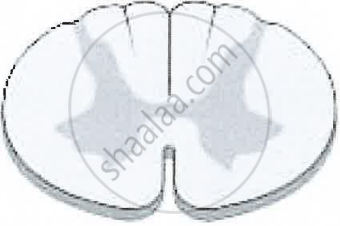
Explain the formation of a typical spinal nerve with the help of a neat labelled diagram.
उत्तर

Formation of Spinal Nerve
- All spinal nerves are of the mixed type i.e. they have some nerve fibre as sensory and some motor.
- Each spinal nerve is formed inside the neural canal of vertebral column by two roots - the posterior or dorsal sensory root and anterior or ventral root.
- Anterior root receives the sensory nerve from the dorsal root ganglion (cell bodies of sensory neurons are located in the ganglion), while the anterior/ventral root gives out the motor nerve.
- The dorsal sensory and the ventral motor nerves together form the mixed spinal nerve.
- It emerges out from both sides of the spinal cord through the intervertebral foramen.
- As soon as it emerges out of vertebral column, it shows three branches viz.
- Ramus dorsalis: From skin and to muscles of dorsal side.
- Ramus ventralis: The largest of the three supplies the organs and muscles on lateral and anterior side.
- Ramus communicans: The smallest of the three and given out from 1st thoracic upto 3rd lumbar (L3) spinal nerve. It joins the sympathetic ganglia.
APPEARS IN
संबंधित प्रश्न
Where is this located?
Ganglia
Give the functions of the spinal cord?
The diagram given below shows the internal structure of the spinal cord, depicting a simple reflex. Study the same and then answer the questions that follow:

(i) Name the parts numbered 1 to 5.
(ii) Using the letters of the alphabet shown in the figure indicate the direction in which an impulse enters and leaves the spinal cord.
(iii) What is the term given to the point of contact between two nerve cells?
(iv) What is meant by ‘simple reflex’ ? Give two examples of simple reflexes and name the stimuli too.
(v) How does the arrangement of nerve cells in the spinal cord differ from that in the brain?
The diagram given below represents the spinal cord of a mammal, seen in a transverse section together with the nerves. Study the diagram and then answer the questions given below:

(i) Label the parts 1—8 indicated by guidelines.
(ii) What do the arrows indicate? What is the pathway indicated termed?
(iii) What type of nerve is shown in the diagram?
Complete the following sentence with appropriate word :
The part of the central nervous system which responds to the reflex action is ________.
Sketch and label T. S. of Spinal cord.
There are ______ pairs of cranial nerves and ______ pairs of spinal nerves.
Which neuron conducts impulse from the spinal cord to the effector organ?
How are cytons and axons arranged in the spinal cord?
Given below is the transverse section of the spinal cord. Read the information below the diagram and fill in the blanks:
The spinal cord extends from the medulla oblongata of the brain and runs down through the whole length of the vertebral column. The spinal cord is covered by the meninges. It conducts impulses from the skin and muscles to the brain. It also conducts impulses from the brain to the muscles of the trunk and limbs.
The spinal cord is a part of the (a) ______ Nervous System. The grey matter in the picture given above consists of (b) ______ while the white matter consists of (c) ______. The spinal cord is concerned with the (d) ______ actions below the neck. (e) ______ is the bony structure that protects the spinal cord.
