Advertisements
Advertisements
प्रश्न
Explain with the help of a suitable diagram conducting system of human heart.
With the help of a neat and labelled diagram, explain the conducting system of human heart.
उत्तर

- Human heart is myogenic (myo-muscle, genic-originating from). The heart beat originates in modified cardiac muscles called Sinoatrial node (SA node) which lies in the wall of right atrium near the opening of superior vena cava.
- The SA node is called pace maker because it has the power to generate of wave of contraction.
- The wave of contraction of cardiac impulse generated by SA node is conducted by cardiac fibres to both the atria causing their contraction (atrial systole).
- The atrioventricular node (AV node) is located in the wall of right atrium near the opening of coronary sinus and receives the wave of contraction generated by the SA node through internodal pathway.
- The Bundle of His arises from AV nodes and divides into right and left bundle branches located in the interventricular septum.
- The bundle branches give rise to Purkinje fibres which penetrate into myocardium of ventricles. The bundle of His and Purkinje fibres conduct the wave of contraction from the AV node to the myocardium of ventricles causing their contraction (ventricular systole).
संबंधित प्रश्न
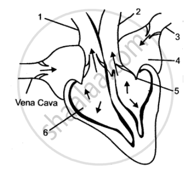
The diagram given below represents the human heart in one phase of its activity. Study the same and then answer the question that follow:
Which part of the heart is contracting in this phase? Give a reason to support your answer.

Name the Following
The valve present between the left atrium and the left ventricle.
State the Location: Semilunar valves of the heart
State the Function: Bundle of His
Column 2 is a list of items related to ideas in Column 1. Match the term in Column 2 with the suitable idea given in Column 1.
| Column 1 | Column 2 |
| (i) Superior vena cava | (a) Collect deoxygenated blood from the wall of the heart. |
| (ii) Inferior vena cava | (b) Carry oxygenated blood to heart muscle. |
| (iii) Pulmonary vein | (c) Collects deoxygenated blood from upper part. |
| (iv) Coronary veins | (d) Collects deoxygenated blood from lower parts. |
| (v) Coronary artery | (e) Brings oxygenated blood from lungs. |
| (vi) Aorta | (f) Large artery |
| (vii) Heart attack | (g) Large vein |
| (viii) Blood Pressure | (h) Oxygenated blood |
| (ix) Tricuspid valve | (i) Sphygmomanometer |
| (x) Bicuspid valve | (j) Allows blood flow from right auricle to right ventricle. |
| (xi) Contraction and relaxation of heart | (k) Blocking of coronary arteries. |
| (l) Cardiac muscle. | |
| (m) Allows blood flow from left auricle to left ventricle. | |
| (n) Allows blood flow from right ventricle of pulmonary aorta. |
Why are the walls of the left ventricle thicker than the other chambers of the heart?
For which of the following reason the SA node acts as the normal pacemaker?
Complete the analogy.
The contraction of heart muscles: Systole:: Relaxation of heart muscles: ______.
______ externally separates ventricles from each other.
The diagram below depicts the human heart in one of its phases. Answer the questions that follow:

- Which part of the heart is in the contraction phase?
- Give a suitable reason to justify your answer in (a).
- Distinguish between Systole and Diastole.
