Advertisements
Advertisements
प्रश्न
Name the following:
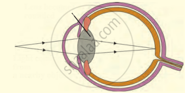
Yellow spot and ciliary muscles are found in.
उत्तर
Eye
APPEARS IN
संबंधित प्रश्न
Which. of the following has normal vision?
(a) Xc Xc
(b) Xc Y
(c) XC Xc
(d) Xc Yc
Name the part of the eye:
which controls the amount of light entering the eye.
Name that part of the eye which is equivalent to the photographic film in a camera.
What shape are your eye-lenses:
when you look at your hand?
Explain why, when it is getting dark at night, it is impossible to make out the colour of cars on the road.
How much is our field of view:
with one eye open?
Fill in the following blank with suitable word:
Having two eyes enables us to judge.................more accurately.
Define the following:
Yellow spot
Give the main function of the following:
Three semicircular canals
Give the main function of the following:
Retina
Answer the following question.
Write the structure of eye lens and state the role of ciliary muscles in the human eye.
What are the functions of tears?
Complete the following sentence with appropriate Word
The part of the human eye where rod cells and cone cells are located is the:
Name the capacity of the eye lens to change its focal length as per need.
Write the name.
The fleshy screen behind cornea.
The following figure show the change in the shape of the lens while seeing distant and nearby objects. Complete the figures by correctly labelling the diagram.

Match the following:
| Column - I | Column - II | ||
| 1 | Retina | a | pathway of light |
| 2 | Pupil | b |
far point comes closer |
| 3 | Ciliary muscles | c |
near point moves away |
| 4 | Myopia | d | screen of the eye |
| 5 | Hypermetropia | e | power of accommodation |
The thin, transparent extension of sclerotic layer found in front of the lens is ______.
Match the following:
| Column - I | Column - II |
| 1. Retina | a. Path way of light |
| 2. Pupil | b. Far point comes closer |
| 3. Ciliary muscles | c. near point moves away |
| 4. Myopia | d. Screen of the eye |
| 5. Hypermetropia | e. Power of accommodation |
Match the terms in column I with those in column II and write down the matching pairs.
| Column I | Column II | ||
| (i) | Conjunctiva | (a) | Viral infection |
| (ii) | Cornea | (b) | Ciliary body |
| (iii) | Choroid | (c) | Spiral-shaped |
| (iv) | Cochlea | (d) | Transparent epithelium |
| (v) | Conjunctivitis | (e) | Suspensory ligament |
| (f) | Contains melanin | ||
| (g) | Transparent but appears black |
