Advertisements
Advertisements
Question
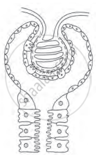
Complete the diagram/chart with correct labels/ information. Write the conceptual details regarding it.

Solution
Malpighian body-

Nephron is the structural and functional unit of kidney.
Malpighian body: Each Malpighian body is about 200μm in diameter and consists of a Bowman’s capsule and glomerulus.
- Glomerulus:
Glomerulus is a bunch of fine blood capillaries located in the cavity of Bowman’s capsule. A small terminal branch of the renal artery, called as afferent arteriole enters the cup cavity (Bowman capsule) and undergoes extensive fine branching to form network of several capillaries. This bunch is called as glomerulus. The capillary wall is fenestrated (perforated). All capillaries reunite and form an efferent arteriole that leaves the cup cavity. The diameter of the afferent arteriole is greater than the efferent arteriole. This creates a high hydrostatic pressure essential for ultrafiltration, in the glomerulus. - Bowman’s capsule:
It is a cup-like structure having double walls composed of squamous epithelium. The outer wall is called as parietal wall and the inner wall is called as visceral wall. The parietal wall is thin consisting of simple squamous epithelium. There is a space called as capsular space / urinary space in between two walls. Visceral wall consists of special type of squamous cells called podocytes having a foot-like pedicel. These podocytes are in close contact with the walls of capillaries of glomerulus. There are small slits called as filtration slits in between adjacent podocytes.
APPEARS IN
RELATED QUESTIONS
Identify the odd one.
Explain the principle of dialysis with the help of a labelled diagram.
Answer the following in short.
List five waste products of plants.
Given below is a set of five terms. Rewrite the terms in their correct order so as to be in logical sequence.
Afferent arteriole, renal vein, secondary capillary network, glomerulus, efferent arteriole.
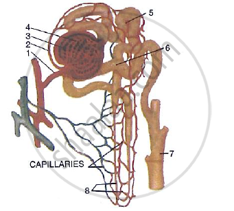
The following diagram represents a mammalian kidney tubule (nephron) and its blood supply.

Parts indicated by the guidelines 1 to 8 are as follows:
1. Afferent arteriole from renal artery
2. efferent arteriole
3. Bowman’s capsule
4. Glomerulus;
5. Proximal convoluted tubule with blood capillaries;
6. Distal convoluted tubule with blood capillaries;
7. collecting tubule;
8. U-shaped loop of Henle
Study the diagram and answer the question that follow:
Where does ultrafiltration take place?
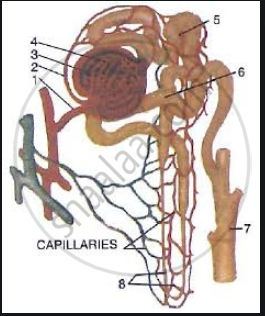
The following diagram represents a mammalian kidney tubule (nephron) and its blood supply.

Parts indicated by the guidelines 1to 8 are as follows:
1. Afferent arteriole from renal artery
2. Efferent arteriole
3. Bowman's capsule
4. Glomerulus
5. Proximal convoluted tubule with blood capillaries
6. Distal convoluted tubule with blood capillaries
7. Collecting tubule
8. U-shaped loop of Henle
Study the diagram and answer the question that follow:
Which structure (normally) contains the lowest concentration of glucose?
Explain the Term: Glomerulus
Complete the following sentence with appropriate word:
The knot of blood vessel inside the Bowman’s capsule is ______.
Write the functional activity of the following structure: Aldosterone
Choose the correct option.
The minor calyx _______.
