Advertisements
Advertisements
Question
Describe the evolutionary change in the pattern of heart among the vertebrates.
Solution 1
- There is only one heart in vertebrates. It is a muscular, hollow organ made of heart muscle fibres. The heart has two different kinds of chambers: ventricles and atria. There are two extra chambers in the heart of lower vertebrates: the sinus venosus and the conus arteriosus, bulbus arteriosus, or truncus arteriosus.
- In higher vertebrates, the persistent parts-the ventricles and auricles-remain throughout development. Nevertheless, these get more intricate when many valves are incorporated within them and they become compartmentalised.
- Fish have a two-chambered heart (one ventricle and one auricle). There are two auxiliary chambers: the sinus venosus and the conus arteriosus. Deoxygenated blood is pumped from the body to the heart via the gills, where it is oxygenated and then returned to the heart. The heart is referred to as a venous heart, and it is called a single circulation.
- The hearts of lungfish, amphibians, and reptiles are composed of three chambers: two auricles and one ventricle. Blood that is oxygenated comes from the gills, lungs, skin, and buccopharyngeal cavity into the left atrium, while blood that is deoxygenated comes from various bodily parts into the right atrium. However, a single ventricle that pumps out mixed blood mixes oxygenated and deoxygenated blood. We refer to this as an incomplete double circulation.
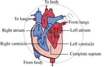
- A complete four-chameled heart (right and left auricles; right and left ventricles) is present in crocodiles, birds, and mammals. Blood that is deoxygenated and oxygenated doesn't mix. While the left side of the heart receives oxygenated blood from the lungs and sends it to other body parts, the right side of the heart receives deoxygenated blood from all other body parts and sends it to the lungs for oxygenation.
- This type of circulation, which combines pulmonary and systemic circulation, is known as full double circulation. In the hearts of birds and mammals, there are no auxiliary chambers.
Solution 2
All vertebrates possess a heart – a hollow muscular organ composed of cardiac muscle fibres. The function of the heart is to pump oxygen to all parts of the body. The evolution of the heart is based on the separation of oxygenated blood from deoxygenated blood for efficient oxygen transport.
In fishes, the heart was like a hollow tube. This evolved into the four-chambered heart in mammals.
Piscean heart
Fish has only two chambers in its heart – one auricle and one ventricle. Since both the auricle and the ventricle remain undivided, only deoxygenated blood passes through it. The deoxygenated blood enters the gills for oxygenation from the ventricle. It has additional chambers such as sinus venosus and conus arteriosus.

Amphibian heart
Amphibians, such as frogs, have three-chambered hearts, with two auricles and one ventricle. The auricle is divided into a right and a left chamber by an inter-auricular septum, while the ventricle remains undivided
Additional chambers such as sinus venosus and conus arteriosus are also present. The oxygenated blood from the lungs enters the left auricle and simultaneously, the deoxygenated blood from the body enters the right auricle. Both these auricles empty into the ventricle, wherein the oxygenated and deoxygenated blood get mixed to some extent.

Reptilian heart
Reptiles have incomplete four-chambered hearts, except for crocodiles, alligators, and gharials. They have only one accessory chamber called sinus venosus. The reptilian heart also shows mixed blood circulation.

Avian and mammalian hearts
They have two pairs of chambers for separating oxygenated and deoxygenated bloods. The heart is divided into four chambers. The upper two chambers are called atria and the lower two chambers are called ventricles. The chambers are separated by a muscular wall that prevents the mixing of the blood rich in oxygen with the blood rich in carbon dioxide.
APPEARS IN
RELATED QUESTIONS
What is the significance of atrio-ventricular node and atrio-ventricular bundle in the functioning of heart?
Conversion of ammonia into uric acid occurs through --------------
A patient’s chart reveals that he has a cardiac output of 7500 mL per minute and a stroke volume of 50 mL. What is his pulse rate (in beats/min)
At any given time there is more blood in the venous system than that of the arterial system. Which of the following features of the veins allows this?
Distinguish between mitral valve and semi lunar valve.
Right ventricular wall is thinner than the left ventricular wall. Why?
What are the heart sounds? When and how are these sounds produced?
Select the correct biological term.
The substances which gives red cells their colour.
Which among the followings is correct during each cardiac cycle?
The cardiac impulse is initiated and conducted further upto ventricle. The correct sequence of conduction of impulse is ______.
Read the following statements and choose the correct option
Statement 1: Atria receive blood from all parts of the body which subsequently flows to ventricles.
Statement 2: Action potential generated at sino-atrial node passes from atria to ventricles.
Define the following terms and give their location?
Purkinje fibre
The walls of ventricles are much thicker than atria. Explain.
Write the features that distinguish between the two.
Open and Closed circulatory system
Answer the following:
What is specific in the heart of crocodiles among reptilians?
