Advertisements
Advertisements
Question
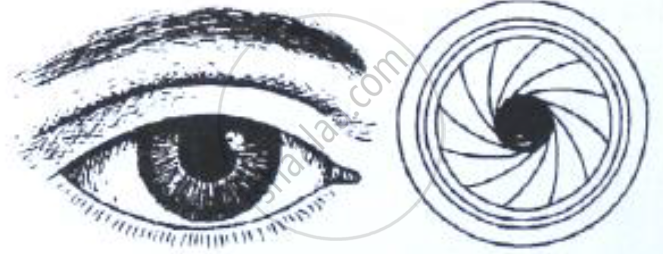
The figure below compares a part of our eye with a part of a photographic camera.

Name the corresponding parts of the eye the camera shown here that are comparable in function.
Solution
Cornea is comparable to the lens cover of the camera. The iris and pupil act like the aperture of a camera.
APPEARS IN
RELATED QUESTIONS
Write short notes on the following: Retina
Out of rods and cones m the retina of your eye:
which work in dim light?
Where does the greatest degree of refraction of light occur in the eye?
What do the ciliary muscles do when you are focusing on a nearby object?
Give the scientific names of the following parts of the eye:
a clear window at the front of the eye.
Note the relationship between the first two words and suggest the suitable word/words for the fourth place.
Cones : Iodopsim :: Rods : ______.
In what two ways is the yellow spot different from the blind spot?
Name the following:
Short sightedness.
Answer the following question.
Trace the sequence of events Whichoccur when a bright light is focused on your eyes.
What is meant by power of accommodation of the eye?
Name the following:
The area where the image is formed but not seen by our eye is termed as.
Give Technical Term:
The cells of the retina that are sensitive to colour.
Mention, if the following statement is True or False
Cones are the receptor cells in the retina of the eye sensitive to dim light.
The following figure show the change in the shape of the lens while seeing distant and nearby objects. Complete the figures by correctly labelling the diagram.

The muscular diaphragm that controls the size of the pupil is ____________.
What kind of lens is there in our eyes? Where does it form the image of an object?
Which cells of the retina enable us to see coloured objects around us?
Vitreous humour is present between ______.
Name the following:
The fibres which collectively hold the lens in position.
Match the terms in column I with those in column II and write down the matching pairs.
| Column I | Column II | ||
| (i) | Conjunctiva | (a) | Viral infection |
| (ii) | Cornea | (b) | Ciliary body |
| (iii) | Choroid | (c) | Spiral-shaped |
| (iv) | Cochlea | (d) | Transparent epithelium |
| (v) | Conjunctivitis | (e) | Suspensory ligament |
| (f) | Contains melanin | ||
| (g) | Transparent but appears black |
