Advertisements
Advertisements
प्रश्न
Describe the structure of the spinal cord.
उत्तर
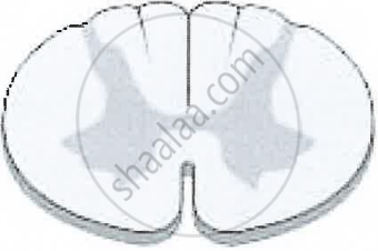
The spinal cord is a cylindrical structure lying in the neural canal of the vertebral column. It is also covered by meninges. It extends from the lower end of the medulla oblongata to the first lumbar vertebra. The posterior-most region of the spinal cord tapers into a thin fibrous thread-like structure called Filum terminate.
Internally, the spinal cord contains a cerebrospinal fluid-filled cavity, known as the central canal. The grey matter of the spinal cord is ‘H’ shaped. The upper end of the letter, ‘H’ forms posterior horns and the lower end forms anterior horns. A bundle of fibres passes into the posterior horn forming the dorsal or afferent root. Fibres pass outward, from the anterior horn forming the ventral or efferent root. These two roots join to form spinal nerves. The white matter is external and has a bundle of nerve tracts. The spinal cord conducts sensory and motor impulses to and from the brain. It controls the reflex actions of the body.
APPEARS IN
संबंधित प्रश्न
Differentiate between following pair with reference to the aspect in bracket.
cerebrum and spinal cord (arrangement of cytons and exons of neurons).
Spinal cord and sympathetic ganglion of autonomous nervous system are connected by ______________.
Give Technical Term:
The fluid which fills the central canal of the spinal cord.
The diagram given below shows the internal structure of the spinal cord, depicting a simple reflex. Study the same and then answer the questions that follow:

(i) Name the parts numbered 1 to 5.
(ii) Using the letters of the alphabet shown in the figure indicate the direction in which an impulse enters and leaves the spinal cord.
(iii) What is the term given to the point of contact between two nerve cells?
(iv) What is meant by ‘simple reflex’ ? Give two examples of simple reflexes and name the stimuli too.
(v) How does the arrangement of nerve cells in the spinal cord differ from that in the brain?
Mention, if the following statement is True or False
Spinal nerves are twelve pair
Sketch and label T. S. of Spinal cord.
How are cytons and axons arranged in the spinal cord?
Explain the formation of a typical spinal nerve with the help of a neat labelled diagram.
Given below is the transverse section of the spinal cord. Read the information below the diagram and fill in the blanks:

The spinal cord extends from the medulla oblongata of the brain and runs down through the whole length of the vertebral column. The spinal cord is covered by the meninges. It conducts impulses from the skin and muscles to the brain. It also conducts impulses from the brain to the muscles of the trunk and limbs.
The spinal cord is a part of the (a) ______ Nervous System. The grey matter in the picture given above consists of (b) ______ while the white matter consists of (c) ______. The spinal cord is concerned with the (d) ______ actions below the neck. (e) ______ is the bony structure that protects the spinal cord.
Mention where in the human body is the following located and state its main function:
Central canal
