Advertisements
Advertisements
प्रश्न
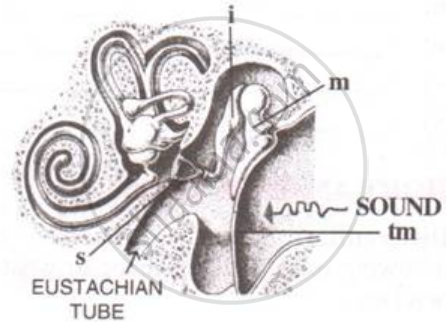
The figure below is the sectional view of a part of the skull showing s sense organ:

Name the sense organ.
उत्तर
Ear
APPEARS IN
संबंधित प्रश्न
Deafness is causal due to the rupturing of the Pinna.
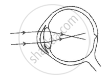
Given below is a diagram depicting a defect of the human eye, study the same and then answer the questions that follow:

(i) Name the defect shown in the diagram.
(ii) What are the two possible that cause this defect?
(iii) Name the type of lens used to correct this defect.
(iv) With the help of a diagram show how the defect shown above is rectified using a suitable lens.
Given in the box below are a set of 14 biological terms. Of these, 12 can be paired into 6 matching pairs. Out of the six pairs, one has been done for you as an example.
Example : endosmosis - Turgid cell.
Identify the remaining five matching pairs :
| Cushing’s syndrome, Turgid cell, Iris, Free of rod and cone cells, Colour of eyes, Hypoglycemia, Active transport, Acrosome, Addison’s disease, Blind spot, Hyperglycemia, Spermatozoa, Endosmosis, Clotting of blood. |
(i) Draw a well labelled diagram of the membranous labyrinth found in the inner ear.
(ii) Based on the diagram drawn above in (i) give a suitable term for each of the following descriptions :
1. The sensory cells that helps in hearing.
2. The part that is responsible for static balance of the body.
3. The membrane covered opening that connects the middle ear to the inner ear.
4. The fluid present in the middle chamber of cochlea.
5. The structure that maintains dynamic equilibrium of the body.
A fluid that occupies the larger cavity of the eye ball behind the lens is
___________.
Differentiate between the Rods and Cones of Retina (Type of pigment)
The ear ossicle which is attached to the tympanum.
Differentiate between members of the following pair with reference to what is asked in bracket.
Dynamic balance and static balance (Definition)
Mention if the following statement is true (T) or false (F) Give reason.
Malleus incus and stapes are collectively called the ear ossicles.
With reference to the human ear, answer the question that follow:
Name the three small bones present in the middle ear. What is the biological term for them collectively ?
With reference to the human ear, answer the question that follow:
Name the nerve, which transmits messages from the ear to the brain.
Draw a labelled diagram of the inner ear.
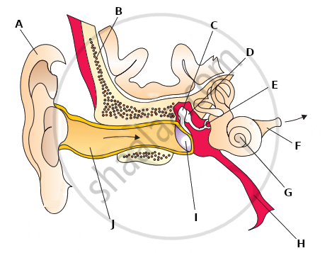
The following diagram refers to the ear of a mammal.

(i) Label the parts 1 to 10 to which the guidelines point.
(ii) Which structure:
(a) converts sound waves into mechanical vibrations?
(b) Converts vibrations into nerve impulses?
(c) Responds to change in position?
(d) Transmits impulses to the brain?
(e) Equalizes atmospheric pressure and pressure in the ear.
State the Function:
Endolymph
Choose the Odd One Out:
List the causes of noise pollution.
Select the option with incorrect identification:

The median canal of cochlea is filled with ______.
Note the relationship between the first two words and suggest the suitable word/words for the fourth place.
Semi-circular canal : Ampulla :: Cochlea : ______.
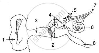
The figure given below shows the principal parts of a human ear. Study the diagram and answer the following questions.
 |
- Label the parts 1 to 8.
- State the role of parts 6, 7 and 8.
- Why is it harmful to use a sharp object to remove ear wax? Mention the number and name of the part involved.
