Advertisements
Advertisements
प्रश्न
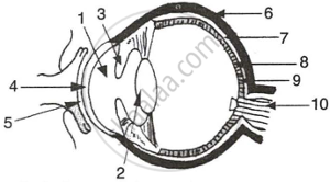
The figure given below refers to the vertical section of the eye of a mammal. Study the figure carefully and answer the following questions.
 |
- Label the guidelines shown as 1 to 10.
- Write one important role of parts shown as 3 and 7.
- Write one structural difference between the parts shown as 9 and 10.
- Mention one functional difference between the parts shown as 6 and 8.
उत्तर
-
- Aqueous Humour
- Lens
- Iris
- Cornea
- Conjunctiva
- Sclera
- Choroid
- Retina
- Fovea centralis
- Optic Nerve
- Iris: The iris has radial and circular muscles that can enlarge or constrict the pupil.
This regulates the amount of light entering the eyes.
Choroid: The choroid includes blood veins that supply the eyeball. - Fovea centralis: The fovea is located in the centre of the macula lutea of the retina.
Optic nerve: The optic nerve connects the eye to the brain, which carries impulses formed by the retina. - Sclera: The sclera is a protective, tough white behind the retina.
Cornea: The cornea is a transparent coating that shields the eye's coloured component and allows light to pass through.
संबंधित प्रश्न
Compare the following: Choroid and retina
Draw labelled diagrams of the following: Eye
How does the eye regulate the amount of light that falls on the retina?
Give the scientific names of the following parts of the eye:
carries signals from an eye to the brain.
Ciliary muscles of human eye can contract or relax. How does it help in the normal functioning of the eye?
An object is moved closer to an eye. What changes must take place in the eye in order to keep the image in sharp focus?
How much is our field of view:
with both eyes open?
Five persons A, B, C, D and E have diabetes, leukaemia, asthma, meningitis and hepatitis, respectively.
Which of these persons can donate eyes?
The animals called predators have:
(a) both the eyes on the sides
(b) one eye on the side and one at the front
(c) one eye on the front and one at the back
(d) both the eyes at the front
The region in the eyes where the rods and cones are located is the
Mention if the following statement is true (T) or false (F) Give reason.
yellow spot of the retina is the region of colour vision
In what two ways is the yellow spot different from the blind spot?
Write whether the following is true or false:
Rods are the receptor cells in the retina of the eye sensitive to dimlight.
Choose the correct answer :
The rods and cones of a vertebrate retina function is to _________
State the Function:
Vitreous humour
Write an Explanation.
Power of accommodation
Match the following
| 1. | Conjunctiva | a. | Coloured part of eye |
| 2. | Cornea | b. | Photosensitive layer |
| 3. | Iris | c. | Refraction |
| 4. | Retina | d. | Protection |
Explain the role of the part of human eye responsible for power of accommodation of human eye.
Name the following:
Place of best vision in the retina of the eye.
Where is the following located?
Yellow spot
