Advertisements
Advertisements
प्रश्न
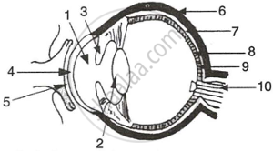
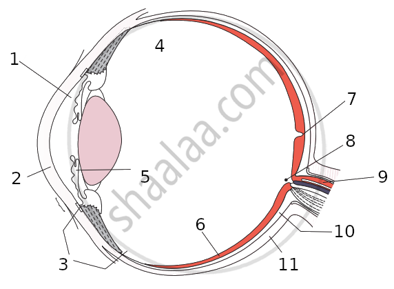
The figure given below refers to the vertical section of the eye of a mammal. Study the figure carefully and answer the following questions.
 |
- Label the guidelines shown as 1 to 10.
- Write one important role of parts shown as 3 and 7.
- Write one structural difference between the parts shown as 9 and 10.
- Mention one functional difference between the parts shown as 6 and 8.
उत्तर
-
- Aqueous Humour
- Lens
- Iris
- Cornea
- Conjunctiva
- Sclera
- Choroid
- Retina
- Fovea centralis
- Optic Nerve
- Iris: The iris has radial and circular muscles that can enlarge or constrict the pupil.
This regulates the amount of light entering the eyes.
Choroid: The choroid includes blood veins that supply the eyeball. - Fovea centralis: The fovea is located in the centre of the macula lutea of the retina.
Optic nerve: The optic nerve connects the eye to the brain, which carries impulses formed by the retina. - Sclera: The sclera is a protective, tough white behind the retina.
Cornea: The cornea is a transparent coating that shields the eye's coloured component and allows light to pass through.
संबंधित प्रश्न
Name the part of the eye:
which controls the amount of light entering the eye.
Name two types of cells in the retina of an eye which respond to light.
What changes take place in the shape of eye-lens:
when the eye is focused on a near object?
There are two types of light-sensitive cells in the human eye:
What is each type called?
What are rods and cones in the retina of an eye? Why is our night vision relatively poor compared to the night vision of an owl?
The size of the pupil of the eye is adjusted by:
(a) cornea
(b) ciliary muscles
(c) optic nerve
(d) iris
Among animals, the predators (like lions) have their eyes facing forward at the front of their heads, whereas the animals of prey (like rabbit) usually have eyes at the sides of their head. Why is this so?
With reference to the functioning of the eye, answer the question that follow:
Name the two structure in the eye responsible for bringing about the change in the shape of the lens.
Give the main function of the following:
Lachrymal glands
Name the following:
The opening through which light enters the eyes.
Name the following:
The most sensitive region of the retina.
A small hole of changing diameter at the centre of Iris is called _______.
Write the name.
The part of human eye that transmits electrical signals to the brain.
Write the name.
The fleshy screen behind cornea.
Match the following
| 1. | Conjunctiva | a. | Coloured part of eye |
| 2. | Cornea | b. | Photosensitive layer |
| 3. | Iris | c. | Refraction |
| 4. | Retina | d. | Protection |
Select the option with incorrect identification:

Which cells of the retina enable us to see coloured objects around us?
Match the following:
| Column - I | Column - II | ||
| 1 | Retina | a | pathway of light |
| 2 | Pupil | b |
far point comes closer |
| 3 | Ciliary muscles | c |
near point moves away |
| 4 | Myopia | d | screen of the eye |
| 5 | Hypermetropia | e | power of accommodation |
Assertion (A): Rods and Cones are photoreceptors in the sclera of eyeball.
Reason (R): Rods are sensitive to dim light.
State the functions of the following:
Iris
