Advertisements
Advertisements
Question
Draw a labeled diagram of the front view of human eye.
Solution

APPEARS IN
RELATED QUESTIONS
Explain, why a normal eye is not able to see distinctly the objects placed closer than 25 cm, without putting any strain on the eye.
How is the amount of light entering the eye controlled?
Ciliary muscles of human eye can contract or relax. How does it help in the normal functioning of the eye?
What are rods and cones in the retina of an eye? Why is our night vision relatively poor compared to the night vision of an owl?
Which of the following changes occur when you walk out of bright sunshine into a poorly lit room?
(a) the pupil becomes larger
(b) the lens becomes thicker
(c) the ciliary muscle relaxes
(d) the pupil becomes smaller
What shape are your eye-lenses:
when you look at your hand?
How much is our field of view:
with one eye open?
Differentiate between members of the following pair with reference to what is asked in bracket.
Rod and cone cells (pigment contained)
Mention if the following statement is true (T) or false (F) Give reason.
Short-sightedness and hyperopia are one and the same thing
Given below is a set of five parts. Rewrite them in correct sequence.
Conjunctiva, retina, cornea, optic nerve, lens.
Give scientific reason:
We cannot clearly see an object kept at a distance less than 25 cm from the eye.
Name the respective organs in which the following are located and mention the main function of each:
(i) Iris
(ii) Semicircular canals
Choose the correct answer.
Colour is detected by ____________
In human eye the part which allows light to enter into the eye is ______.
Match the following:
| Column - I | Column - II | ||
| 1 | Retina | a | pathway of light |
| 2 | Pupil | b |
far point comes closer |
| 3 | Ciliary muscles | c |
near point moves away |
| 4 | Myopia | d | screen of the eye |
| 5 | Hypermetropia | e | power of accommodation |
Assertion (A): Rods and Cones are photoreceptors in the sclera of eyeball.
Reason (R): Rods are sensitive to dim light.
Write in proper sequence the names of all the parts of the human eye through which the light rays coming from an object pass before they form an image on the retina.
Name the following:
Capacity of the eye to focus at different distances.
With reference to human eye answer the question that follow:
Name the part of the eye associated with the regulation of the size of pupil.
The figure given below refers to the vertical section of the eye of a mammal. Study the figure carefully and answer the following questions.
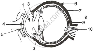 |
- Label the guidelines shown as 1 to 10.
- Write one important role of parts shown as 3 and 7.
- Write one structural difference between the parts shown as 9 and 10.
- Mention one functional difference between the parts shown as 6 and 8.
