Advertisements
Advertisements
Question
The figure below is the sectional view of a part of the skull showing a sense organ:
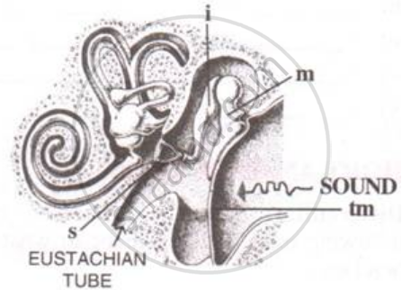
What do you call the part shown in the form of a spiral? What is its function?
Solution
Cochlea. The vibrating movements in the hair of the sense cells of cochlea transmit the impulse for hearing to the brain via auditory nerve.
APPEARS IN
RELATED QUESTIONS
Explain the following:
Mechanism through which a sound produces a nerve impulse in the inner ear.
Photoreceptor cells are present in __________.
(A) blind spot
(B) retina
(C) cochlea
(D) cornea
The number of oscillations per second of a vibrating object is called its time period.
Mention the exact location of the Eustachian tube
Name the following:
The ear ossicle attached to the tympanum.
State whether the following statements are true (T) or false (F). If false, correct them by changing any one single word in each.
Semi-circular canals are concerned with static (positional) balance.
Given below is certain structure. Write against them its functional acivity.
organ of corti and ……………….
Name that part of ear which vibrates when outside sound falls on it.
Name the nerve which carries electrical impulses from the cochlea of ear to the brain.
Give the main function of the following:
Cochlea
The figure below is the sectional view of a part of the skull showing a sense organ:

What are the parts labeled 'm', 'i' and 's'? What do these parts constitute collectively?
The range of audible frequency for the human ear is
Describe the structure and function of the human ear?
The median canal of cochlea is filled with ______.
Which of the following structures equalises the air pressure on either side of the tympanum?
Define the following term:
Ear ossicles
With reference to human ear answer the question that follow:
Name the part of the ear associated with the amplification of vibrations.
- Draw a neat and well labelled diagram of the membranous labyrinth found in the inner ear.
- Based on the diagram drawn above in (i), give a suitable term for each of the following descriptions:
- The structure responsible for hearing.
- The sensory cells that help in hearing.
- The membrane-covered opening that connects the middle ear to inner ear.
- The nerves that carry impulses from the ear to the brain.
- The tube which equalises the air pressure on either side of the ear drum.
The figure given below shows the principal parts of a human ear. Study the diagram and answer the following questions.
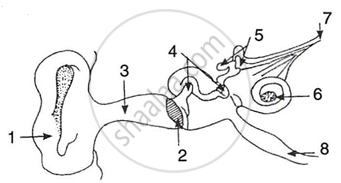 |
- Label the parts 1 to 8.
- State the role of parts 6, 7 and 8.
- Why is it harmful to use a sharp object to remove ear wax? Mention the number and name of the part involved.
