Advertisements
Advertisements
प्रश्न
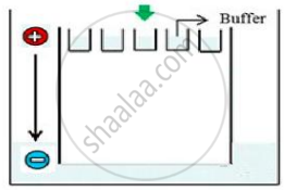
Carefully observe the given picture. A mixture of DNA with fragments ranging from 200 base pairs to 2500 base pairs was electrophoresed on agarose gel with the following arrangement.

(a) What result will be obtained on staining with ethidium bromide? Explain with reason.
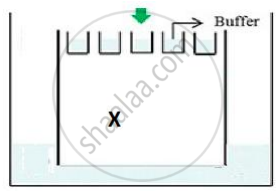
(b) The above setup was modified and a band with 250 base pairs was obtained at X.

What change(s) were made to the previous design to obtain a band at X? Why did the band appear at position X?
उत्तर
(a) No bands will be obtained as all DNA will be seen in the well only; DNA fragments being negatively charged will not move towards the negative end/cathode. DNA being negatively charged will remain stationed at the positive end/anode end of the agar block.
(b)
- The position of the positive terminal/end/anode and the negative terminal/end/cathode was inter-changed.
- The fragment with the least base pairs will get separated faster and move faster to the anode end.
APPEARS IN
संबंधित प्रश्न
Explain with the help of a suitable example the naming of a restriction endonuclease.
Name and describe the technique that helps in separating the DNA fragments formed by the use of restriction endonuclease
Make a chart (with diagrammatic representation) showing a restriction enzyme, the substrate DNA on which it acts, the site at which it cuts DNA and the product it produces.
Explain the roles of the following with the help of an example each in recombinant DNA technology :
Restriction Enzymes
Answer the following question.
Write the use of restriction endonuclease in the formation of recombinant DNA.
DNA fragments separate according to size through?
DNA strands on a gel stained with ethidium bromide when viewed under UV radiation, appear as ______
A specific recognition sequence identified by endonucleases to make cuts at specific positions within the DNA is ______
Given below is the restriction site of a restriction endonuclease Pst-I and the cleavage sites on a DNA molecule.
\[\ce{5' C - T - G - C - A \overset{\downarrow}{-}{G 3'}}\]
\[\ce{3' G\underset{\uparrow}{-} A - C - G - T - C 5'}\]
Choose the option that gives the correct resultant fragments by the action of the enzyme Pst-I.
How are DNA fragments visualised once they are separated by gel electrophoresis?
