Advertisements
Advertisements
प्रश्न
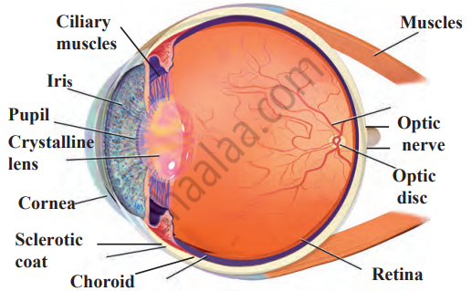
Draw a neat and labelled diagram of the structure of the human eye.
उत्तर

संबंधित प्रश्न
Distinguish between: blind spot and yellow spot
Explain, why a normal eye is not able to see distinctly the objects placed closer than 25 cm, without putting any strain on the eye.
What kind of lens is present in the human eye?
Give the scientific names of the following parts of the eye:
a clear window at the front of the eye.
Fill in the following blank with suitable word:
Most of the refraction of light rays entering the eye occurs at the outer surface of the.............
Fill in the following blank with suitable word:
The iris controls the amount of................entering the eye.
What are rods and cones in the retina of an eye? Why is our night vision relatively poor compared to the night vision of an owl?
After testing the eyes of a child, the optician has prescribed the following lenses for his spectacles:
Left eye : + 2.00 D
Right eye : + 2.25 D
The child is suffering from the defect of vision called:
(a) short-sightedness
(b) long-sightedness
(c) cataract
(d) presbyopia
How much is our field of view:
with both eyes open?
Having two eyes gives a person:
(a) deeper field of view
(b) coloured field of view
(c) rear field of view
(d) wider field of view
Name the following:
The photoreceptors found in the retina of the eye.
Differentiate between members of the following pair with reference to what is asked in bracket.
Rod and cone cells (pigment contained)
Mention if the following statement is true (T) or false (F) Give reason.
Blind spot is called so because no image is formed on it.
With reference to the functioning of the eye, answer the question that follow:
Name the cells of the retina and its respective pigments which get activated in the dark.
State the function of each of the following parts of the human eye:
(i) Cornea
(ii) Iris
(iii) Pupil
(iv) Retina
Millions of people of the developing countries are suffering from corneal blindness. This disease can be cured by replacing the defective cornea with the cornea of a donated eye. Your school has organised a campaign in the school and its neighbourhood in order to create awareness about this fact and motivate people to donate their eyes after death. How can you along with your classmates contribute in this noble cause? State the objectives of organising such campaigns in schools.
What is the function of iris and the muscles connected to the lens in human eye?
Define the following:
Yellow spot
Define the following:
Blind spot
Choose the correct answer.
Aperture controlling passage to light into the eye is ___________
What are the functions of tears?
Differentiate between:
Vitreous humour and Aqueous humour.
Choose the Odd One Out:
Choose the Odd One Out:
Complete the following sentence with appropriate Word
The part of the human eye where rod cells and cone cells are located is the:
Write the name.
The part of human eye that transmits electrical signals to the brain.
For a healthy human eye, the distant point is infinite distance.
Why the human eye is compared with camera?
The pigmented circular area seen in front of the eye:
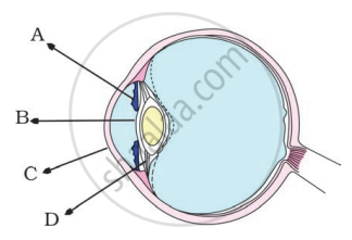
In the figure of the human eye, the cornea is represented by the letter

What kind of lens is there in our eyes? Where does it form the image of an object?
Write down the names of parts of the eye in the blank spaces shown in the figure.

In human eye the part which allows light to enter into the eye is ______.
Match the following:
| Column - I | Column - II |
| 1. Retina | a) Path way of light |
| 2. Pupil | b) Far point comes closer |
| 3. Ciliary muscles | c) near point moves away |
| 4. Myopia | d) Screen of the eye |
| 5. Hypermetropia | e) Power of accommodation |
An aperture that controls the passage of light into the eye is ______.
Which of the following is responsible for the adjustment of the size of pupil?
Write the main functional activity of the following structure.
Choroid
Write the main functional activity of the following structure.
Ciliary body and suspensory ligament
With reference to human eye answer the question that follow:
Name the part of the eye associated with the regulation of the shape of lens.
