Advertisements
Advertisements
Question
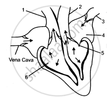
The figure below represents the internal structure of a mammalian heart and the associated blood vessels.
(i) (a) Name each of the structures labeled 1, 2, 3, 4, 5, 6, 7 and 8.
(b) State the function of each of the structures 5, 6, 7, and 8.
(ii) (a) State the function of the heart as an entire organ.
(b) Why are the walls of the left ventricle more muscular than the right?
Solution
(i) (a)
1. Right auricle,
2. Left ventricle,
3. Pulmonary artery,
4. Pulmonary vein,
5. Inferior vena cava,
6. Aorta,
7. Pulmonary semilunar valve,
8. Tricuspid valve.
(b)
5 — carry deoxygenated blood from the body parts to the right auricle of the heart.
6 — distributes blood all over the body.
7 — prevents the backflow of the blood into the ventricles.
8 — prevents the reverse flow of the blood from the right ventricle into the right auricle,
(ii) (a) The heart makes the blood to circulate all over the body.
(b) The walls of the left ventricle are more muscular because they have to pump blood to a larger distance than the right so that walls have to withstand high pressure.
APPEARS IN
RELATED QUESTIONS
Name the following:
The inflammation of pericardium.
Choose the correct answer:
In mammals the opening of post caval in the right auricle is guarded by _________
The diagram given below represents the human heart in one phase of its activity. Study the same and then answer the question that follow:
Name the parts numbered 1 to 6.

Explain the Term
Electrocardiogram (ECG)
What is the importance of valves in the heart?
______ guards the opening of inferior vena cava.
Left atrium : pulmonary vein : right ventricle : ____________.
Heart is made up of ______.
Draw the neat Labelled diagram of the internal structure of the human heart.
Give scientific reason.
Human heart is called as myogenic and autorhythmic.
