English

Human Heart

Advertisements

Topics

  • Structure of the Heart
  • Layers of the Heart Wall
  • Chambers of the Heart
  • Valves of the Heart
  • Pathway of Blood through the Heart

Structure of the Heart:

The heart is located almost at the centre of the thoracic cage. It is situated behind the ribs, between the two lungs, and slightly inclined to the left side.

  • The size of our heart is approximately equal to one's own fist, and it weighs about 360 grams. It is covered by a double-layered peritoneal membrane.
  • A fluid is present between the two membranes, which protects the heart from friction and mechanical shocks.
  • The human heart is a muscular organ composed of involuntary cardiac muscles. These muscles contract and relax with a definite rhythm, known as the beating of the heart.

The human heart is a four-chambered muscular organ, about the size and shape of a closed fist. Two-thirds of the heart's mass is positioned to the left of the midline of the body. The heart is enclosed in a pericardial sac, providing protection and support. The sac is lined with the parietal layer of a serous membrane. The visceral layer of this serous membrane forms the epicardium, which is the outer layer of the heart.

Structure of Heart and Blood Circulation

Layers of the Heart Wall:

  1. Epicardium (Outer Layer): The epicardium is the outermost layer of the heart wall. It serves as a protective layer and is made up of connective tissue. It also contains blood vessels and nerves that supply the heart.
  2. Myocardium (Middle Layer): The myocardium is the thickest and most muscular layer of the heart wall. It is composed of cardiac muscle tissue that contracts to pump blood. This layer is responsible for the heart's powerful beating action.
  3. Endocardium (Inner Layer): The endocardium is the innermost layer of the heart wall. It is a thin, smooth layer that lines the inside of the heart chambers. It helps to prevent blood from sticking to the heart walls and keeps the heart functioning smoothly.

Chambers of the Heart:

The internal cavity of the heart is divided into four chambers:

  1. Right atrium
  2. Right ventricle
  3. Left atrium
  4. Left ventricle

The atria are the upper, thin-walled chambers of the heart. They receive blood from veins: the right atrium gets deoxygenated blood from the body, and the left atrium gets oxygenated blood from the lungs.

The ventricles are the lower, thick-walled chambers of the heart. They pump blood forcefully: the right ventricle sends deoxygenated blood to the lungs, and the left ventricle pumps oxygenated blood to the entire body.

The thickness of the chamber walls varies because thicker walls (more myocardium) are needed to generate more force for pumping blood.

Valves of the Heart:

The heart has valves to ensure blood flows in one direction. There are two main types of valves: atrioventricular valves and semilunar valves.

1. Atrioventricular Valves (Cuspid Valves)

  • Located between the atria and ventricles.
  • The right atrioventricular valve is the tricuspid valve.
  • The left atrioventricular valve is the bicuspid or mitral valve.

2. Semilunar Valves

  • Located at the base of the large vessels leaving the ventricles.
  • The pulmonary semilunar valve is between the right ventricle and the pulmonary trunk.
  • The aortic semilunar valve is between the left ventricle and the aorta.

When the ventricles contract, atrioventricular valves close to prevent blood from flowing backward into the atria. When the ventricles relax, semilunar valves close to prevent blood from flowing back into the ventricles.

Pathway of Blood through the Heart:

The heart has two sides that work together, pumping blood simultaneously. Blood flows from the right atrium to the right ventricle, which pumps it to the lungs for oxygen. Oxygenated blood then moves from the lungs to the left atrium, then to the left ventricle, which pumps it to the rest of the body.

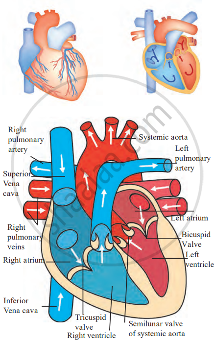
Blood Circulation Pathway Through the Heart and Body
 
Blood Supply to the Myocardium:

The myocardium, the heart's muscle layer, needs a constant supply of oxygen and nutrients. The right and left coronary arteries deliver oxygen-rich blood to the myocardium. After the blood delivers oxygen and collects waste, it flows through cardiac veins, which mostly empty into the coronary sinus. The coronary sinus then drains into the right atrium.

If you would like to contribute notes or other learning material, please submit them using the button below.

Video Tutorials

We have provided more than 1 series of video tutorials for some topics to help you get a better understanding of the topic.

Series 1


Series 2


Series 3


Shaalaa.com | Arteries and veins of the heart

Shaalaa.com


Next video


Shaalaa.com


Arteries and veins of the heart [00:05:54]
S
Series: Arteries and veins of the heart
0%


Advertisements
Share
Notifications

Englishहिंदीमराठी


      Forgot password?
Use app×