Advertisements
Advertisements
Question
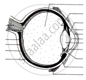
Write the function of the human eye and label parts of the figure given below.

Solution
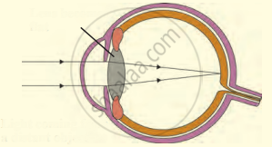
- Double convex transparent crystalline lens: It provides small adjustments of the focal length to focus the image. This lens creates a real and inverted image of an object on the screen inside the eye.
- Optic nerve: The sensitive cells on the retina get excited when light falls on them and generate electric signals. The optic nerve carries these signals to the brain.
- Retina: It is the screen inside the human eye which contains light-sensitive cells. Image of an object is formed on this screen.
- Pupil: The pupil contracts or widens depending on the intensity of light incident on the eye.
APPEARS IN
RELATED QUESTIONS
List the parts of the human eye that control the amount of light entering into it. Explain how they perform this function.
The human eye can focus objects at different distances by adjusting the focal length of the eye lens. This is due to ______.
Differentiate between: Rods and cones
Myopia is an example of ______.
With the help of ciliary muscles the human eye can change its curvature and thus alter the focal length of its lens. State the changes that occur in the curvature and focal length of the eye lens while viewing (a) a distance object, (b) nearby objects.
A person got his eyes tested by an optician. The prescription for the spectacle lenses to be made reads :
Left eye : +2.50 D
Right eye : +2.00 D
State whether these lenses are thicker in the middle or at the edges.
Out of rods and cones m the retina of your eye:
which detect colour?
What changes the shape of lens in the eye?
What is the:
near point of a normal human eye?
Fill in the following blank with suitable word:
The part of eye which alters the size of the pupil is............
There are two types of light-sensitive cells in the human eye:
To what is each type of cell sensitive?
Draw a simple diagram of the human eye and label clearly the cornea, iris, pupil, ciliary muscles, eye-lens, retina, optic nerve and blind spot.
How does the eye adjust itself to deal with light of varying intensity?
The human eye forms the image of an object at its ______.
Which part brings the image into sharp focus on the retina? How does it do this?
When is a person said to have developed cataract in his eye? How is the vision of a person having cataract restored?
Mention if the following statement is true (T) or false (F) Give reason.
Blind spot is called so because no image is formed on it.
Given below is certain structure. Write against them its functional acivity.
retina and ……………………….
Give scientific reason:
One can sense colours only in bright light.
Define the following:
Yellow spot
Label the following diagram :

Name the respective organs in which the following are located and mention the main function of each:
(i) Iris
(ii) Semicircular canals
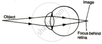
The diagram alongside represents a certain defect of vision of the human eye.
(i) Name the defect.
(ii) Describe briefly the condition in the eye responsible for the defect.
(iii) Redraw the figure by adding a suitable lens correcting the defect. Label the parts through which light-rays pass.
(iv) What special advantage do human beings derive in having both eyes facing forward?

Name the following:
The focal length of the lens is altered by the contraction of which type of muscles.
Mention, if the following statement is True or False
The least distance of distinct vision for the human eye is 25 cm
A small hole of changing diameter at the centre of Iris is called _______.
Write the name.
The fleshy screen behind cornea.
Write the name.
The screen with light sensitive cells in human eye.
Write an Explanation.
Farthest distance of distinct vision
The following figure show the change in the shape of the lens while seeing distant and nearby objects. Complete the figures by correctly labelling the diagram.

______ is the structural and functional unit of living organisms.
The larynx has fold of tissue which vibrate with the passage of air to produce sound.
When light rays enter the eye, most of the refraction occurs at the ____________.
From where the following nerves arise.
Optic nerve
______ is a transparent layer.
Which one of the following statements is NOT correct?
Match the following:
| Column - I | Column - II |
| 1. Retina | a) Path way of light |
| 2. Pupil | b) Far point comes closer |
| 3. Ciliary muscles | c) near point moves away |
| 4. Myopia | d) Screen of the eye |
| 5. Hypermetropia | e) Power of accommodation |
Match the following:
| Column - I | Column - II |
| 1. Retina | a. Path way of light |
| 2. Pupil | b. Far point comes closer |
| 3. Ciliary muscles | c. near point moves away |
| 4. Myopia | d. Screen of the eye |
| 5. Hypermetropia | e. Power of accommodation |
Name the following:
The circular opening enclosed by iris.
Give reason:
Blind spot is considered as 'area of no vision'.
