Advertisements
Advertisements
प्रश्न
Complete the following sentence with appropriate Word
The aperture in the eye through which light enters is the:
विकल्प
Pupil
Conjunctiva
Ciliary muscles
Choroid
उत्तर
Pupil
APPEARS IN
संबंधित प्रश्न
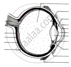
Draw a neat and labelled diagram of the structure of the human eye.
What do the ciliary muscles do when you are focusing on a nearby object?
Draw a simple diagram of the human eye and label clearly the cornea, iris, pupil, ciliary muscles, eye-lens, retina, optic nerve and blind spot.
Nocturnal animals (animals which sleep during the day and come out at night) tend to have wide pupils and lot of rods in their retinas. Suggest reasons for this.
Label the following diagram :

Differentiate between:
Vitreous humour and Aqueous humour.
State the Function:
Choroid coat in the eye
State the Function:
Visual purple
Draw a scientifically correct labelled diagram of a human eye and answer the questions based on it:
- Name the type of lens in the human eye.
- Name the screen at which the maximum amount of incident light is refracted?
- State the nature of the image formed of the object on the screen inside the eye.
The larynx has fold of tissue which vibrate with the passage of air to produce sound.
The pigmented circular area seen in front of the eye:
With reference to human eye, answer the following question.
What is blind spot?
| Column I | Column II | ||
| 1 | Retina | a | Path way of light |
| 2 | Pupil | b | Far point comes closer |
| 3 | Ciliary muscles | c | near point moves away |
| 4 | Myopia | d | Screen of the eye |
| 5 | Hypermetropia | e | Power of accomodation |
Match the following.
| Column - I | Column - II | ||
| 1 | Retina | a | Path way of light |
| 2 | Pupil | b | Far point comes closer |
| 3 | Ciliary muscles | c | near point moves away |
| 4 | Myopia | d | screen of the eye |
| 5 | Hypermetropia | e | Power of accomadation |
State the functions of the following:
Ciliary muscles
Match the terms in column I with those in column II and write down the matching pairs.
| Column I | Column II | ||
| (i) | Conjunctiva | (a) | Viral infection |
| (ii) | Cornea | (b) | Ciliary body |
| (iii) | Choroid | (c) | Spiral-shaped |
| (iv) | Cochlea | (d) | Transparent epithelium |
| (v) | Conjunctivitis | (e) | Suspensory ligament |
| (f) | Contains melanin | ||
| (g) | Transparent but appears black |
Write the main functional activity of the following structure.
Choroid
Write the main functional activity of the following structure.
Ciliary body and suspensory ligament
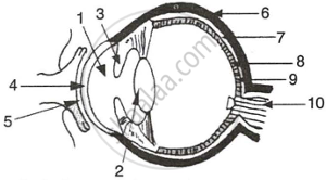
The figure given below refers to the vertical section of the eye of a mammal. Study the figure carefully and answer the following questions.
 |
- Label the guidelines shown as 1 to 10.
- Write one important role of parts shown as 3 and 7.
- Write one structural difference between the parts shown as 9 and 10.
- Mention one functional difference between the parts shown as 6 and 8.
