Topics
Sexual Reproduction in Flowering Plants
- Flower - a Fascinating Organ of Angiosperms
- Parts of Flower
- Accessory Organs
- Essential Parts of Flower: Androecium
- Essential Parts of Flower: Gynoecium
- Sexual Reproduction in Flowering Plants
- Pre-fertilisation in Flowering Plant: Structures and Events
- Development of Anther
- Transverse Section of Mature Anther (Microsporangium)
- Microsporogenesis
- Microspores and Pollen Grains
- Development of Male Gametophyte
- Advantages and Disadvantages of Pollen Grains
- Structure of Ovule (Megasporangium)
- Types of Ovules
- Megasporogenesis
- Development of Female Gametophyte or Embryo Sac
- Pollination
- Outbreeding Devices
- Artificial Hybridization
- Kinds of Pollination
- Self Pollination (Autogamy)
- Cross Pollination
- Agents of Pollination
- Abiotic Agents
- Biotic Agents
- Fertilization Process
- Fertilization Process
- Post Fertilisation in Plant: Structures and Events
- Development of Endosperm
- Post Fertilization in Plant: Development of Embryo (Embryogeny)
- Development of Seed
- Development of Fruit
- Apomixis
- Polyembryony
Reproduction in Organisms
- Life Span of Organisms
- Maximum Life Span of Organisms
- Reproduction in Organisms
- Types of Reproduction
- Asexual Reproduction
- Sexual Reproduction in Animals
- Asexual Reproduction in Plant
- Asexual Reproduction in Animal
- Budding
- Vegetative Reproduction
- Natural Vegetative Reproduction
- Artificial Vegetative Reproduction
- Artificial Vegetative Reproduction
- Artificial Vegetative Reproduction
- Fission
- Budding
- Sporulation (Sporogenesis)
- Fragmentation
- Different Phases in Sexual Reproduction
- Sexual Reproduction in Animals
- Pre-fertilisation Events in Organisms
- Fertilisation in Organisms
- Post-fertilisation Events in Organisms
Reproduction
Genetics and Evolution
Human Reproduction
Reproductive Health
Biology and Human Welfare
Environmental Issues
- Environmental Issues
- Prevention of Air Pollution
- Controlling Vehicular Air Pollution: a Case Study of Delhi
- Introduction of Water Pollution and Its Control
- Effects of Domestic Sewage and Industrial Effluents on Water
- A Case Study of Integrated Waste Water Treatment
- Solid Wastes
- Agrochemicals and Their Effects
- Radioactive Wastes
- Greenhouse Effect and Climate Change
- Ozone Depletion in the Stratosphere
- Degradation by Improper Resource Utilisation and Maintenance
- Deforestation and Its Causes
- Radioactive Waste Management and E-waste
- Solid Waste Management
- Noise Pollution
- Environmental Issues
Biotechnology
Principles of Inheritance and Variation
- Introduction of Principles of Inheritance and Variation
- Mendelism
- Terminology Related to Mendelism
- Mendel’s experiments on pea plant
- Monohybrid Cross
- Gregor Johann Mendel – Father of Genetics
- Punnett Square
- Back Cross and Test Cross
- Mendelian Inheritance - Mendel’s Law of Heredity
- The Law of Dominance
- The Law of Segregation (Law of Purity of Gametes)
- The Law of Independent Assortment
- Intragenic Interactions - Incomplete Dominance
- Intragenic Interactions - Codominance
- Gregor Johann Mendel – Father of Genetics
- Extensions of Mendelian Genetics (Deviation from Mendelism)
- Intragenic Interactions - Incomplete Dominance
- Intragenic Interactions - Dominance
- Intragenic Interactions - Codominance
- Multiple Alleles
- Intragenic Interactions - Pleiotropy
- Polygenic Inheritance
- Chromosomal Theory of Inheritance
- Historical Development of Chromosome Theory
- Comparison Between Gene and Chromosome Behaviour
- Chromosomal Theory of Inheritance: Law of Segregation
- Chromosomal Theory of Inheritance: Law of Independent Assortment
- Linkage and Recombination
- Sex Determination
- Sex Determination in Some Insects
- Sex Determination in Human
- Sex Determination in Birds
- Sex Determination in Honey Bees
- Concept of Mutation
- Pedigree Analysis
- Genetic Disorders
- Mendelian Genetics
- Chromosomal Abnormalities
- Heredity and Variation
- Linkage and Crossing Over
- Principles of Inheritance and Variation Question
Molecular Basis of Inheritance
- Introduction of Molecular Basis of Inheritance
- Deoxyribonucleic Acid (DNA) and Its Structure
- Structure of Polynucleotide Chain
- Packaging of DNA Helix
- Search for Genetic Material
- Introduction of Search for Genetic Material
- The Genetic Material is a DNA
- Properties of Genetic Material (DNA Versus RNA)
- The RNA World
- DNA Replication
- The Experimental Proof
- The Machinery and the Enzymes
- Protein Synthesis
- Introduction of Transcription
- Transcription Unit
- Transcription Unit and the Gene
- Types of RNA and the Process of Transcription
- Genetic Code
- Genetic Code
- Genetic Code
- tRNA – the Adapter Molecule
- Translation
- Regulation of Gene Expression
- Operon Concept
- Human Genome Project
- DNA Fingerprinting Technique
- Structure of DNA and RNA
- Structure of Nucleotide
- Rice Genome Project
- Molecular Basis of Inheritance (Questions)
Ecology
Evolution
- Origin and Evolution of Universe and Earth
- Theories of Origin of Life
- Evolution of Life Forms - a Theory
- Evidences for Biological Evolution
- Theories of Biological Evolution
- Adaptive Radiation
- Organic Evolution
- Hardy Weinberg’s Principle
- Brief Account of Evolution
- Human Evolution
- Darwinism
- Micro and Macro Evolution
- Speciation
- Evolution Stages
- Modern Synthetic Theory of Evolution
- Gene Flow and Genetic Drift
- Evolution
Human Health and Diseases
- Introduction of Human Health and Diseases
- Common Diseases in Human Beings
- Immunity
- Types of Immunity
- Vaccination and Immunization
- Allergies (Hypersensitivity)
- Autoimmunity
- Human Immune System
- Sexually Transmitted Diseases (STD)
- Cancer
- Introduction of Drugs and Alcohol Abuse
- Drugs and Alcohol Abuse
- Adolescence - Drug and Alcohol Abuse
- Addiction and Dependence
- Effects of Drug and Alcohol
- Prevention and Control of Drugs and Alcohol Abuse
- Infectious and Non Infectious Disease
- Maintaining Good Health, Yoga, Excercise
- Human Health and Diseases (Questions)
Strategies for Enhancement in Food Production
Microbes in Human Welfare
- Microbes in Human Welfare
- Microbes in Household Products
- Microbes in Industrial Production
- Microbes in Sewage Treatment
- Microbes in Production of Biogas
- Microbes as Biocontrol Agents
- Microbes as Biofertilizers
- Energy Generation
- Production and Judicious Use
- Microbes in Human Welfare
Biotechnology - Principles and Processes
- Process and Principles of Biotechnology
- Restriction Enzymes
- Cloning Vectors
- Competent Host (For Transformation with Recombinant DNA)
- Processes of Recombinant DNA Technology
Biotechnology and Its Application
Organisms and Populations
- Introduction of Organisms and Populations
- Ecology (Organism, Population, Community and Biome)
- Introduction of Organisms and Environment
- Major Abiotic Factors
- Responses to Abiotic Factors
- Population Attributes
- Population Growth
- Life History Variation
- Population Interactions
- Population and Ecological Adaptations
- Organisms and Populations (Questions)
Ecosystem
- Ecosystem
- Introduction and Types of Ecosystem
- Ecosystem - Structure and Function
- Productivity
- Decomposition
- Concept of Energy Flow in an Ecosystem
- Ecological Pyramids
- Ecological Succession
- Nutrient Cycles
- Ecosystem Services
- Ecosystems Patterns
Biodiversity and Its Conservation
- Biodiversity
- Species on Earth and Species in India
- Patterns of Biodiversity
- Importance of Species Diversity to the Ecosystem
- Loss of Biodiversity
- Conservation of Biodiversity
- Endangered Organisms
- Importance of Biodiversity
- Extinction
- Red Data Book
- Biodiversity and Its Conservation (Questions)
- Lymphoid organs:
- Primary or central lymphoid organs: Thymus gland, Bone marrow
- Secondary lymphoid organs: Lymph node, Spleen, Peyer’s patches, Tonsils, Spleen, Adenoids, MALT, GALT, BALT.
Notes
Lymphoid organs:
- The immune system of an organism consists of several structurally and functionally different organs and tissues that are widely dispersed in the body.
- The organs involved in the origin, maturation, and proliferation of lymphocytes are called lymphoid organs.
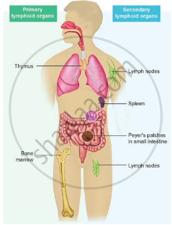
Lymphoid organs in the human body
- Based on their functions, they are classified into primary or central lymphoid organs and secondary or peripheral lymphoid organs.
- The primary lymphoid organs provide an appropriate environment for lymphocytic maturation.
- The secondary lymphoid organs trap antigens and make it available for mature lymphocytes, which can effectively fight against these antigens.
Notes
1) Primary lymphoid organs:
- The Bursa of Fabricius of birds, bone marrow, and thymus gland of mammals constitute the primary lymphoid organs involved in the production and early selection of lymphocytes.
- These lymphocytes become dedicated to a particular antigenic specificity.
- Only when the lymphocytes mature in the primary lymphoidal organs, they become immunocompetent cells.
- In mammals, B cell maturation occurs in the bone marrow and T cell maturation occurs in the thymus.
i) Thymus:
- The thymus is a flat and bilobed organ located behind the sternun, above the heart.
- Each lobe of the thymus contains numerous lobules, separated from each other by connective tissue called septa.
- Each lobule is differentiated into two compartments, the outer compartment or outer cortex is densely packed with immature T cells called thymocytes, whereas the inner compartment or medulla is sparsely populated with mature thymocytes.
- One of its main secretions is the hormone thymosin. It stimulates the T cell to become mature and immunocompetent.
- By the early teens, the thymus begins to atrophy and is replaced by adipose tissue. Thus thymus is most active during the neonatal and pre-adolescent periods.

Primary lymphoid organ - Thymus A) Location B) Structure
ii) Bone marrow:
- Bone marrow is a lymphoid tissue found within the spongy portion of the bone.
- Bone marrow contains stem cells known as haematopoietic cells.
- These cells have the potential to multiply through cell division and either remain as stem cells or differentiate and mature into different kinds of blood cells.
Notes
2) Secondary or peripheral lymphoid organs:
- In secondary or peripheral lymphoid organs, antigen is localized so that it can be effectively exposed to mature lymphocytes.
- The best examples are lymph nodes, appendix, Peyer’s patches of gastrointestinal tract, tonsils, adenoids, spleen, MALT (Mucosal-Associated Lymphoid Tissue), GALT (Gut-Associated Lymphoid Tissue), BALT (Bronchial/Tracheal-Associated Lymphoid Tissue).
i) Lymph node:
- Lymph node is a small bean-shaped structure and is part of the body’s immune system.
- It is the first one to encounter the antigen that enters the tissue spaces.
- Lymph nodes filter and trap substances that travel through the lymphatic fluid.
- They are packed tightly with white blood cells, namely lymphocytes and macrophages.
- There are hundreds of lymph nodes found throughout the body. They are connected to one another by lymph vessels.
- Lymph is a clear, transparent, colourless, mobile, and extracellular fluid connective tissue.
- As the lymph percolates through the lymph node, the particulate antigen brought in by the lymph will be trapped by the phagocytic cells, follicular and interdigitating dendritic cells.
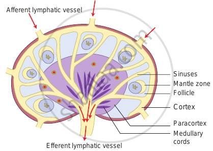
Secondary lymphoid organ – Structure of lymph node
- The lymph node has three zones.
- They are the cortex, paracortex, and medulla.
- The outermost layer of the lymph node is called cortex, which consists of B-lymphocytes, macrophages, and follicular dendritic cells.
- The paracortex zone is beneath the cortex, which is richly populated by T lymphocytes and interdigitating dendritic cells.
- The innermost zone is called the medulla which is sparsely populated by lymphocytes, but many of them are plasma cells, which actively secrete antibody molecules.
- As the lymph enters, it slowly percolates through the cortex, paracortex, and medulla, giving sufficient chance for the phagocytic cells and dendritic cells to trap the antigen brought by the lymph.
- The lymph leaving a node carries enriched antibodies secreted by the medullary plasma cells against the antigens that enter the lymph node.
- Sometimes visible swelling of lymph nodes occurs due to active immune response and increased concentration of lymphocytes. Thus swollen lymph nodes may signal an infection. There are several groups of lymph nodes.
- The most frequently enlarged lymph nodes are found in the neck, under the chin, in the armpits, and in the groin.
ii) Peyer’s patches:
- Peyer’s patches are oval-shaped areas of thickened tissue that are embedded in the mucus-secreting lining of the small intestine of humans and other vertebrate animals.
- Peyer’s patches contain a variety of immune cells, including macrophages, dendritic cells, T cells, and B cells.
iii) Tonsils:
- The tonsils (palatine tonsils) are a pair of soft tissue masses located at the back of the throat (pharynx).
- The tonsils are part of the lymphatic system, which helps to fight infections. They stop invading germs including bacteria and viruses.
iv) Spleen:
- The spleen is a secondary lymphoid organ located in the upper part of the abdominal cavity close to the diaphragm.
- The spleen contains B and T cells. It brings humoral and cell mediated immunity.
v) Adenoids:
- The adenoids are glands located in the roof of the mouth, behind the soft palate where the nose connects to the throat.
- The adenoids produce antibodies that help to fight infections.
- Typically, the adenoids shrink during adolescence and may disappear by adulthood.
vi) Mucosa-associated lymphoid tissue (MALT):
- The mucosa-associated lymphoid tissue (MALT) is a diffuse system of small concentrations of lymphoid tissue in the alimentary, respiratory, and urino-genital tracts.
- MALT is populated by lymphocytes such as T and B cells, as well as plasma cells and macrophages, each of which is well situated to encounter antigens passing through the mucosal epithelium.
- It also possesses IgA antibodies.
vii) Gut-associated lymphoid tissue (GALT):
- Gut-associated lymphoid tissue (GALT) is a component of the mucosa-associated lymphoid tissue (MALT) which works in the immune system to protect the body from invasion in the gut.
viii) Bronchus Associated Lymphoid Tissues (BALT):
- Bronchus Associated Lymphoid Tissues (BALT) also a component of MALT is made of lymphoid tissue (tonsils, lymph nodes, lymph follicles) is found in the respiratory mucosae from the nasal cavities to the lungs.
If you would like to contribute notes or other learning material, please submit them using the button below.
