Advertisements
Advertisements
Question
What is the function of iris and the muscles connected to the lens in human eye?
Solution
- Function of Iris: The iris is a muscular diaphragm that controls the size of the pupil, which, in turn, controls the amount of light entering the eye. It also gives color to the eye.
- Function of ciliary muscles: The ciliary muscles hold the eye lens in position and adjust its focal length by expanding and contracting.
APPEARS IN
RELATED QUESTIONS
Write the function of retina in human eye.
The human eye can focus objects at different distances by adjusting the focal length of the eye lens. This is due to ______.
Distinguish between: aqueous humor and vitreous humor
Which. of the following has normal vision?
(a) Xc Xc
(b) Xc Y
(c) XC Xc
(d) Xc Yc
With the help of ciliary muscles the human eye can change its curvature and thus alter the focal length of its lens. State the changes that occur in the curvature and focal length of the eye lens while viewing (a) a distance object, (b) nearby objects.
Out of rods and cones m the retina of your eye:
which detect colour?
State whether the following statement is true or false:
The image formed on our retina is upside-down
Why does it take some time to see objects in a dim room when you enter the room from bright sunshine outside?
There are two types of light-sensitive cells in the human eye:
What is each type called?
Explain why, we cannot see our seats first when we enter a darkened cinema hall from bright light but gradually they become visible.
Refraction of light in the eye occurs at:
(a) the lens only
(b) the cornea only
(c) both the cornea and the lens
(d) the pupil
What shape are your eye-lenses:
when you look at a distant tree?
What is presbyopia? Write two causes of this defect. Name the type of lens which can be used to correct presbyopia.
Five persons A, B, C, D and E have diabetes, leukaemia, asthma, meningitis and hepatitis, respectively.
Which of these persons cannot donate eyes?
Having two eyes gives a person:
(a) deeper field of view
(b) coloured field of view
(c) rear field of view
(d) wider field of view
Name the respective organs in which the following are located and mention the main function of each:
(i) Iris
(ii) Semicircular canals
Choose the correct answer.
In the chemistry of vision, the photosensitive substance is _________________
Differentiate between:
Vitreous humour and Aqueous humour.
Name the following:
Yellow spot and ciliary muscles are found in.
Choose the Odd One Out:
Write the name.
The screen with light sensitive cells in human eye.
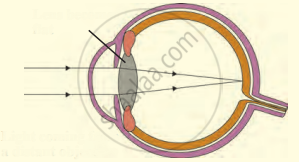
The following figure show the change in the shape of the lens while seeing distant and nearby objects. Complete the figures by correctly labelling the diagram.

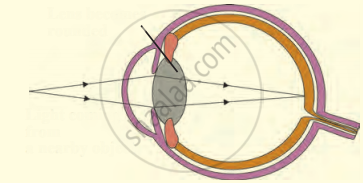
The following figure show the change in the shape of the lens while seeing distant and nearby objects. Complete the figures by correctly labelling the diagram.

______ is the structural and functional unit of living organisms.
The pigmented circular area seen in front of the eye:
A student sitting on the last bench can read the letters written on the blackboard but is not able to read the letters written in his text book. Which of the following statements is correct?
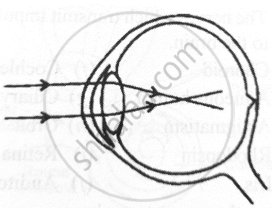
Given below is a diagram depicting a defect of the human eye. Answer the questions that follow:

- Give the scientific term for the defect.
- Mention one possible reason for the defect.
- What type of lens can be used to correct the defect?
From where the following nerves arise.
Optic nerve
With reference to human eye, answer the following question.
What is aqueous humor?
With neat, labeled diagram describe the structure of retina of eye.
State the functions of the following:
Iris
Name the following:
Place of best vision in the retina of the eye.
Name the following:
The layer of the wall of the eye-ball that corresponds to the black lining of the box of a camera
Name the following:
Two pigments of the sensory cells.
Name the following:
Three layers of the eye ball.
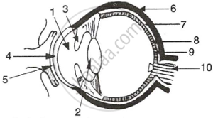
The figure given below refers to the vertical section of the eye of a mammal. Study the figure carefully and answer the following questions.
 |
- Label the guidelines shown as 1 to 10.
- Write one important role of parts shown as 3 and 7.
- Write one structural difference between the parts shown as 9 and 10.
- Mention one functional difference between the parts shown as 6 and 8.
