Topics
The Living World
- Introduction of the Living World
- What is ‘Living’?
- Diversity in the Living World
- Taxonomic Hierarchy of Living Organisms: Unit of Classification
- Taxonomical Aids
Class 11
Class 12
Biological Classification
- Introduction of Biological Classification
- History of Classification
- Five Kingdom Classification
- Kingdom Monera
- Division of Kingdom Monera
- Examples of Kingdom Monera
- Bacteria
- Classification of Bacteria
- Structure of Bacteria
- Life Processes in Bacteria
- Reproduction in Bacteria
- Economic Importance of Bacteria
- Kingdom Protista
- Kingdom Protista
- Protozoa
- Kingdom Fungi
- Division of Kingdom Fungi
- Fungi
- Classification of Fungi
- Reproduction in Fungi
- Economic Importance of Fungi
- Examples of Fungi
- Classification of Kingdom Plantae
- Kingdom Animalia
- Viruses, Viroids, Prions and Lichens
Plant Kingdom
Animal Kingdom
- Kingdom Animalia
- Criteria for New System of Classification
- Classification of Kingdom Animalia
- Invertebrata and Vertebrata
- Non Chordates (Invertebrata)
- Phylum: Porifera
- Phylum: Cnidaria/Coelenterata
- Phylum: Ctenophora
- Phylum: Platyhelminthes
- Phylum: Aschelminthes
- Phylum: Annelida
- Phylum: Arthropoda
- Phylum: Mollusca
- Phylum: Echinodermata
- Phylum: Hemichordata
- Phylum: Chordata
- Subphylum -Vertebrata/Craniata
- Class: Cyclostomata
- Class: Chondrichthyes
- Class: Osteichthyes
- Class: Amphibia
- Class: Reptilia
- Class: Aves
- Class: Mammalia
Morphology of Flowering Plants
- Plant Morphology
- Root System
- The Leaf
- Shoot System
- The Inflorescence
- The Flower
- Parts of Flower
- The Fruit
- The Seed
- Classification and Structure of Seeds
- Structure of a Dicotyledonous Seed
- Structure of Monocotyledonous Seed
- Semi-technical Description of a Typical Flowering Plant
- Plant Forms and Functions
- Description of Some Important Families
Anatomy of Flowering Plants
- Anatomy and Functions of Different Parts of Flowering Plants
- Tissues - “The Teams of Workers”
- Plant and Animals Tissue
- Plant Tissues
- Meristems or Meristematic Tissues
- Permanent Tissue
- Simple Permanent Tissues (Supporting Tissue)
- Complex Permanent Tissues
- Complex Permanent Tissue: Xylem Structure and Function (Conducting Tissue)
- Complex Permanent Tissue: Phloem Structure and Function (Conducting Tissue)
- Tissue System
- Epidermal Tissue System
- Ground Tissue System
- Vascular Tissue System
- Anatomy of Dicotyledonous and Monocotyledonous Plants
- Dicotyledonous Root
- Monocotyledonous Root
- Dicotyledonous Stem
- Monocotyledonous Stem
- Isobilateral (Monocotyledonous) Leaf
- Dorsiventral (Dicotyledonous) Leaf
- Secondary Growth
- Vascular Cambium
- Cork Cambium
- Secondary Growth in Roots
Structural Organisation in Animals
- Introduction of Structural Organisation in Animals
- Tissues - “The Teams of Workers”
- Animal Tissues
- Epithelial Tissue
- Connective Tissue
- Muscular Tissue
- Neural Tissues
- Earthworm - Lampito Mauritii
- Morphology of Earthworm
- Anatomy of Earthworm
- Cockroach - Periplaneta Americana
- Morphology of Cockroach
- Anatomy of Cockroach
- Frog - Rana Hexadactyla
- Morphology of Frog
- Anatomy of Frog
Cell: the Unit of Life
- Cell: Structural and Functional Unit of Life
- The Invention of the Microscope and the Discovery of Cell
- Cell Theory
- Overview of Cell
- Organisms Show Variety in Cell Number, Shape and Size
- Prokaryotic Cells
- Cell Envelope and Its Modifications
- Ribosomes and Inclusion Bodies
- Structure of Prokaryotic and Eukaryotic Cells
- Eukaryotic Cells
- Cell Membrane
- Cell Wall
- Endomembrane System
- Mitochondria
- Plastids
- Ribosomes
- Cilia and Flagella
- Centrosome and Centrioles
- Cytoskeleton
- Nucleus
- Microbodies
- Plant Cell and Animal Cell
- Structure and Functions of Cell Envelope, Cell Membrane, Cell Wall, Cell Organelles
Biomolecules
- Biomolecules
- How to Analyse Chemical Composition?
- Primary and Secondary Metabolites
- Biomacromolecules
- Proteins
- Polysaccharides
- Biomolecules in the Cell
- Nucleic Acids
- Structure of Proteins
- Nature of Bond Linking Monomers in a Polymer
- Dynamic State of Body Constituents – Concept of Metabolism
- Metabolic Basis for Living
- The Living State
- Enzymes - Chemical Reactions
- Enzymes - High Rates of Chemical Conversions
- Nature of Enzyme Action
- Factors Affecting Enzyme Activity
- Classification and Nomenclature of Enzymes
- Enzymes - Co-factors
- Carbohydrates
- Structure and Function of Lipids
- Carbohydrates
Cell Cycle and Cell Division
- Cell Cycle
- Phases of Cell Cycle
- M Phase
- Karyokinesis (Nuclear Division)
- Cytokinesis
- Significance of Mitosis
- Meiosis as a Reduction Division
- Stages of Meiosis: Meiosis I
- Stages of Meiosis: Meiosis II
- Significance of Meiosis
Transport in Plants
- Introduction of Transport in Plants
- Means of Transport in Plants
- Simple Diffusion
- Facilitated Diffusion
- Active Transport
- Concept of Osmosis
- Turgidity and Flaccidity (Plasmolysis)
- Concept of Imbibition
- Comparison of Different Transport Processes
- Plant Water Relation
- Water Potential (ψ)
- Long Distance Transport of Water
- Plants Absorb Water
- Water Movement up a Plant
- Transpiration
- Transpiration - Transpiration and Photosynthesis – a Compromise
- Uptake and Transport of Mineral Nutrients
- Uptake of Mineral Ions
- Transport of Mineral Ions
- Phloem Transport - Flow from Source to Sink
- Phloem Transport - Pressure Flow Or Mass Flow Hypothesis
- Diffusion of Gases
- Structure of Stomatal Apparatus
Mineral Nutrition
- Plant Mineral Nutrition
- Methods to Study the Mineral Requirements of Plants
- Essential Mineral Elements
- Criteria for Essentiality
- Macro and Micro Nutrients and Their Role
- Deficiency Symptoms of Essential Elements
- Toxicity of Micronutrients
- Mechanism of Absorption of Elements
- Soil as Reservoir of Essential Elements
- Biological Nitrogen Fixation
- Nitrogen Cycle
Photosynthesis in Higher Plants
- Introduction of Photosynthesis in Higher Plants
- What Do We Know?
- Early Experiments on Photosynthesis
- Where Does Photosynthesis Take Place?
- Pigments Are Involved in Photosynthesis
- Light Dependent Reaction (Hill Reaction \ Light Reaction)
- Electron Transport
- Electron Transport - Photolysis / Splitting of Water
- Electron Transport - Cyclic and Non-cyclic Photo-phosphorylation
- Electron Transport - Chemiosmotic Hypothesis
- ATP and NADPH Used
- Primary Acceptor of CO2
- The Calvin Cycle
- The C4 Pathway
- Photorespiration
- Factors Affecting Photosynthesis
Respiration in Plants
- Introduction of Respiration in Plants
- Plants Breathe
- Types of Respiration: Aerobic and Anaerobic Respiration
- Phases of Respiration: Glycolysis
- Phases of Respiration: Fermentation
- Aerobic Respiration
- Phases of Respiration: Tricarboxylic Acid Cycle (Citric Acid Cycle Or Kreb’s Cycle)
- Phases of Respiration: Electron Transport Chain (Electron Transfer System)
- Phases of Respiration: Electron Transport System (Ets) and Oxidative Phosphorylation
- Respiratory Balance Sheet
- Amphibolic Pathways
- Respiratory Quotient (R.Q.)
Plant Growth and Development
- Introduction of Plant Growth and Development
- Growth in Plants
- Plant Growth Generally is Indeterminate
- Plant Growth is Measurable
- Phases of Plant Growth
- Plant Growth Rate
- Conditions Necessary for Plant Growth
- Differentiation, Dedifferentiation and Redifferentiation
- Concept of Development
- Plant Growth Regulators
- Characteristics of Growth Regulators
- Discovery of Plant Growth Regulators
- Physiological Effects of Plant Growth Regulators
- Photoperiodism
- Vernalisation
- Formation of Seed and Fruit
Digestion and Absorption
- Introduction of Digestion and Absorption
- Alimentary Canal
- Digestive Glands
- Role of Digestive Enzymes and Gastrointestinal Hormones
- Peristalsis, Digestion, Absorption and Assimilation of Proteins, Carbohydrates and Fats
- Calorific Values of Proteins
- Calorific Values of Carbohydrates
- Calorific Values of Fats
- Digestion of Food
- Absorption of Digested Products
- Nutritional and Digestive Tract Disorders
- Egestion of Food
- Nutritional and Digestive Tract Disorders
Breathing and Exchange of Gases
- Introduction of Breating and Exchange of Gases
- Respiratory Organs
- Human Respiratory System
- Mechanism of respiration-Breathing
- Respiratory Volumes and Capacities
- Exchange of Gases
- Transport of Gases - Transport of Oxygen
- Transport of Gases - Transport of Carbon Dioxide
- Regulation of Breathing / Respiration
- Disorders of Respiratory System
Body Fluids and Circulation
- Introduction of Body Fluids and Circulation
- Blood
- Composition of Blood: Plasma (The Liquid Portion of Blood)
- Composition of Blood: Red Blood Cells (Erythrocytes)
- Composition of Blood: White Blood Cells (Leukocytes)
- Composition of Blood: Blood Platelets (Thrombocytes)
- Blood Transfusion and Blood Groups (ABO and Rh system)
- Function of Platelets - Clotting of Blood (Coagulation)
- Lymph and Lymphatic System
- Types of Closed Circulation
- Blood Circulatory System in Human
- Human Heart
- Blood Vessels
- Circulatory Pathways
- Cardiac Cycle
- Cardiac Output
- Heart Beat - Heart Sounds "LUBB" and "DUP"
- Electrocardiograph (ECG)
- Types of Closed Circulation
- Regulation of Cardiac Activity
- Disorders of Circulatory System
Excretory Products and Their Elimination
- Introduction of Excretory Products and Their Elimination
- Modes of Excretion: Ammonotelism, Ureotelism, Uricotelism
- Human Excretory System
- Function of the Kidney - “Production of Urine”
- Function of the Tubules
- Mechanism of Concentration of the Filtrate
- Regulation of Kidney Function
- Micturition
- Accessory Excretory Organs
- Common Disorders of the Urinary System
Locomotion and Movement
- Introduction of Locomotion and Movement
- Types of Movement
- Muscles
- Structure of Contractile Proteins
- Mechanism of Muscle Contraction
- Skeletal System
- The Human Skeleton: Axial Skeleton
- The Human Skeleton: Appendicular Skeleton
- Joints and Its Classification
- Disorders of Muscular and Skeletal System
Neural Control and Coordination
- Introduction of Neural Control and Coordination
- Neural Tissue
- Neuron (Or Nerve Cell) and Its Types
- Neuron (Or Nerve Cell) and Its Types
- Generation and Conduction of Nerve Impulse
- Human Nervous System
- Major Division of the Nervous System
- Central Nervous System (CNS)
- The Human Brain - Forebrain
- The Human Brain - Forebrain
- The Spinal Cord
- Peripheral Nervous System (PNS)
- Reflex and Reflex Action
- Reflex Arc
- Sense Organs
- Human Eye
- Working of the Human Eye
- Human Ear
Chemical Coordination and Integration
- Introduction of Chemical Coordination and Integration
- Human Endocrine Glands
- The Hypothalamus
- Pituitary Gland or Hypophysis Gland
- The Pineal Gland
- Thyroid Gland
- Parathyroid Gland
- Thymus Gland
- Adrenal Gland (Suprarenal Gland)
- Pancreas (Islets of Langerhans)
- Testis
- Ovary
- Hormones of Heart, Kidney and Gastrointestinal Tract
- Mechanism of Hormone Action
- Role of Hormones as Messengers and Regulators
- Hypo and Hyperactivity and Related Disorders
Reproduction in Organisms
- Life Span of Organisms
- Maximum Life Span of Organisms
- Reproduction in Organisms
- Types of Reproduction
- Asexual Reproduction
- Sexual Reproduction in Animals
- Asexual Reproduction in Plant
- Budding
- Agamospermy
- Vegetative Reproduction
- Natural Vegetative Reproduction
- Artificial Vegetative Reproduction
- Artificial Vegetative Reproduction
- Artificial Vegetative Reproduction
- Asexual Reproduction in Animal
- Fission
- Budding
- Sporulation (Sporogenesis)
- Fragmentation
- Regeneration
- Different Phases in Sexual Reproduction
- Sexual Reproduction in Animals
- Pre-fertilisation Events in Organisms
- Fertilisation in Organisms
- Post-fertilisation Events in Organisms
- Parthenogenesis
- Advantages and Disadvantages of Parthenogenesis
Sexual Reproduction in Flowering Plants
- Flower - a Fascinating Organ of Angiosperms
- Whorls of Flower
- Sexuality in Flowers
- Plant Sex
- Flower Symmetry
- Parts of Flower
- Accessory Organs
- Essential Parts of Flower: Androecium
- Essential Parts of Flower: Gynoecium
- Sexual Reproduction in Flowering Plants
- Pre-fertilisation in Flowering Plant: Structures and Events
- Development of Anther
- Transverse Section of Mature Anther (Microsporangium)
- Microsporogenesis
- Microspores and Pollen Grains
- Development of Male Gametophyte
- Advantages and Disadvantages of Pollen Grains
- Structure of Ovule (Megasporangium)
- Types of Ovules
- Megasporogenesis
- Development of Female Gametophyte or Embryo Sac
- Pollination
- Kinds of Pollination
- Self Pollination (Autogamy)
- Cross Pollination
- Agents of Pollination
- Abiotic Agents
- Biotic Agents
- Outbreeding Devices
- Artificial Hybridization
- Fertilization Process
- Fertilization Process
- Post Fertilisation in Plant: Structures and Events
- Development of Endosperm
- Post Fertilization in Plant: Development of Embryo (Embryogeny)
- Development of Seed
- Development of Fruit
- Apomixis
- Polyembryony
Human Reproduction
- Human Reproduction
- Human Reproduction
- The Male Reproductive System
- Testes
- Accessory Ducts
- Accessory Glands
- External Genitalia
- The Female Reproductive System
- Ovaries
- Accessory Ducts
- External Genitalia (Vulva)
- Accessory Glands
- Mammary Glands
- Gametogenesis
- Spermatogenesis
- Oogenesis
- Menstrual Cycle (Ovarian Cycle)
- Menstrual Hygiene
- Fertilization and Implantation
- Fertilization in Human
- Embryonic Development in Human
- Implantation in Human
- Pregnancy and Embryonic Development
- Parturition and Lactation
Reproductive Health
Principles of Inheritance and Variation
- Introduction of Principles of Inheritance and Variation
- Mendelism
- Terminology Related to Mendelism
- Mendel’s experiments on pea plant
- Monohybrid Cross
- Punnett Square
- Back Cross and Test Cross
- Mendelian Inheritance - Mendel’s Law of Heredity
- The Law of Dominance
- The Law of Segregation (Law of Purity of Gametes)
- The Law of Independent Assortment
- Gregor Johann Mendel – Father of Genetics
- Extensions of Mendelian Genetics (Deviation from Mendelism)
- Intragenic Interactions - Incomplete Dominance
- Intragenic Interactions - Dominance
- Intragenic Interactions - Codominance
- Multiple Alleles
- Intragenic Interactions - Pleiotropy
- Polygenic Inheritance
- Chromosomal Theory of Inheritance
- Historical Development of Chromosome Theory
- Comparison Between Gene and Chromosome Behaviour
- Chromosomal Theory of Inheritance: Law of Segregation
- Chromosomal Theory of Inheritance: Law of Independent Assortment
- Linkage and Recombination
- Sex Determination
- Sex Determination in Some Insects
- Sex Determination in Human
- Sex Determination in Birds
- Sex Determination in Honey Bees
- Concept of Mutation
- Pedigree Analysis
- Genetic Disorders
- Mendelian Genetics
- Chromosomal Abnormalities
- Linkage and Crossing Over
Molecular Basis of Inheritance
- Introduction of Molecular Basis of Inheritance
- Deoxyribonucleic Acid (DNA) and Its Structure
- Structure of Polynucleotide Chain
- Packaging of DNA Helix
- Search for Genetic Material
- Introduction of Search for Genetic Material
- The Genetic Material is a DNA
- Properties of Genetic Material (DNA Versus RNA)
- The RNA World
- DNA Replication
- The Experimental Proof
- The Machinery and the Enzymes
- Protein Synthesis
- Transcription
- Transcription Unit
- Transcription Unit and the Gene
- Types of RNA and the Process of Transcription
- Genetic Code
- Genetic Code
- Genetic Code
- tRNA – the Adapter Molecule
- Translation
- Mechanism of Translation
- Initiation of Translation
- Elongation of Translation
- Termination of Translation
- Regulation of Gene Expression
- Operon Concept
- Human Genome Project
- Human Genome Project
- Human Genome Project
- Human Genome Project
- Applications and Future Challenges
- DNA Fingerprinting Technique
- Polymorphism
- DNA Fingerprinting Technique
- DNA Fingerprinting Technique
Evolution
- Evolution
- Origin and Evolution of Universe and Earth
- Theories of Origin of Life
- Evolution of Life Forms - a Theory
- Evidences for Biological Evolution
- Theories of Biological Evolution
- Lamarck’s Theory of Evolution
- Darwinism
- Modern Synthetic Theory of Evolution
- Adaptive Radiation
- Organic Evolution
- Hardy Weinberg’s Principle
- Brief Account of Evolution
- Human Evolution
Human Health and Diseases
- Introduction of Human Health and Diseases
- Common Diseases in Human Beings
- Bacterial Diseases
- Viral Diseases
- Protozoan Diseases
- Helminthic Diseases
- Fungal Diseases
- Maintenance of Personal and Public Hygiene
- Immunity
- Types of Immunity
- Innate Immunity
- Acquired Immunity
- Immune Responses
- Vaccination and Immunization
- Organ Transplantation
- Allergies (Hypersensitivity)
- Autoimmunity
- Human Immune System
- Acquired Immuno Deficiency Syndrome (AIDS)
- Cancer
- Drugs and Alcohol Abuse
- Adolescence - Drug and Alcohol Abuse
- Addiction and Dependence
- Effects of Drug and Alcohol
- Prevention and Control of Drugs and Alcohol Abuse
Strategies for Enhancement in Food Production
Microbes in Human Welfare
- Introduction of Microbes in Human Welfare
- Microbes in Household Products
- Microbes in Industrial Production
- Microbes in Sewage Treatment
- Microbes in Production of Biogas
- Microbes as Biocontrol Agents
- Microbes as Biofertilizers
Biotechnology - Principles and Processes
- Introduction of Principles and Processes of Biotechnology
- Biotechnology
- Principles of Biotechnology
- Tools of Recombinant DNA Technology
- Restriction Enzymes
- Cloning Vectors
- Competent Host (For Transformation with Recombinant DNA)
- Processes of Recombinant DNA Technology
Biotechnology and Its Application
Organisms and Populations
- Introduction of Organisms and Populations
- Ecology (Organism, Population, Community and Biome)
- Organism and Its Environment
- Introduction of Organisms and Environment
- Major Abiotic Factors
- Responses to Abiotic Factors
- Population and Ecological Adaptations
- Population Attributes
- Life History Variation
- Population Growth
- Population Interactions
Ecosystem
Biodiversity and Its Conservation
- Introduction to Biodiversity and Conservation
- Biodiversity
- Patterns of Biodiversity
- Importance of Biodiversity
- Red Data Book
- Loss of Biodiversity
- Conservation of Biodiversity
- Biosphere Reserve
- National Park
- Wildlife Sanctuary
Environmental Issues
- Environmental Issues
- Pollution
- Air Pollution and Its Causes
- Sources of Air Pollution
- Prevention of Air Pollution
- Controlling Vehicular Air Pollution: a Case Study of Delhi
- Water Pollution and Its Causes
- Sources of Water Pollution
- Effects of Domestic Sewage and Industrial Effluents on Water
- A Case Study of Integrated Waste Water Treatment
- Prevention of Water Pollution
- Solid Wastes
- Agrochemicals and Their Effects
- Radioactive Wastes
- Greenhouse Effect and Climate Change
- Ozone Depletion in the Stratosphere
- Degradation by Improper Resource Utilisation and Maintenance
- Deforestation and Its Causes
- Consequences of Deforestation
- Forest Conservation
- Case Study of People's Participation in Conservation of Forests
- Testes
- Histology of seminiferous tubules
Notes
Testes:
|
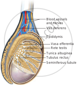
Diagrammatic view of male reproductive system (part of testis is open to show inner details) |
- Testes are the primary male sex organs.
- They are a pair of ovoid bodies lying in the scrotum.
- The testes are situated outside the abdominal cavity within a pouch called the scrotum.
- Since viable sperms cannot be produced at normal body temperature, the scrotum is placed outside the abdominal cavity to provide a temperature 2-3°C lower than the normal internal body temperature. Thus, the scrotum acts as a thermoregulator for spermatogenesis.
- In adults, each testis is oval in shape, with a length of about 4 to 5 cm and a width of about 2 to 3 cm.

Testis showing inner details
- Testes are enclosed in an outer tough capsule of collagenous connective tissue, the tunica albuginea.
- Each testis is divided by septa into about 200 - 250 lobules called testicular lobules.
- The scrotum is connected to the abdominal cavity through a passage termed inguinal-canal. Through this canal, the testis descends down into the scrotal sacs at the time of birth. The spermatic cord in males passes through the inguinal canal.
- Testes are covered by three coats.
- Tunica Vaginalis: The outermost covering is called Tunica vaginalis which has a parietal and visceral layer. It covers the whole testis except its posterior border from where the testicular vessels and nerves enter the testis.
- Tunica albuginea: The middle tough capsule of collagenous connective tissue called tunica albuginea. The Tunica albuginea is a dense, white fibrous coat covering the testis all around. The posterior border tunica albuginea is thickened to form a vertical septum called the Mediastinum testis.
- Tunica Vasculosa: Tunica vasculosa is the innermost vascular coat of the testis lining testicular lobules.
- The failure of one or both testes to descend down into the scrotal sacs is known as cryptorchism (crypto – hidden + orchis – testicle). It occurs in 1 – 3 percent of new born males. A surgical correction at a young age can rectify the defect, else these individuals may become sterile and are unable to produce viable sperms. It can also lead to cancer.
- Internally scrotum is lined by dartos muscle and spermatic fascia. Dartos muscle helps in the regulation of the temperature within the scrotum during the cold season, it becomes contracted in cold & during the warm season, it becomes relaxed. Cremaster muscles line inside the wall of the scrotal & inguinal canal region and help in the elevation of testes.
- Each testis is attached to the walls of the scrotal sac through flexible, elastic fibres. This group of fibres is called Gubernaculum or Mesorchium.
- Each testis is attached to the dorsal body wall of the abdominal cavity through a cord termed the spermatic cord. This cord is made up of elastin fibres & spermatic fascia. The contents of cord are vas deferens, gonadal veins, gonadal arteries, nerves and lymphatics.
Notes
Histology of seminiferous tubules:
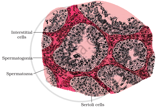
- Each lobule contains 1-3 highly coiled testicular tubules or seminiferous tubules. These highly convoluted tubules which form 80 per cent of the testicular substance are the sites for sperm production.
- The stratified epithelium of the seminiferous tubule is made of two types of cells namely sertoli cells or nurse cells and spermatogonic cells or male germ cells.
- Sertoli cells are elongated and pyramidal and provide nourishment to the sperms till maturation. They also secrete inhibin, a hormone which is involved in the negative feedback control of sperm production. Spermatogonic cells divide meiotically and differentiate to produce spermatozoa.
- The regions outside the seminiferous tubules called interstitial spaces contain:
1) Small blood vessels
2) Interstitial cells or Leydig cells
3) Other immunologically competent cells - Leydig cells synthesise and secrete testicular hormones called androgens, also called male sex hormones.
- Interstitial cells or Leydig cells are embedded in the soft connective tissue surrounding the seminiferous tubules. These cells are endocrine in nature and secrete androgens (also called male sex hormones) namely the testosterone hormone which initiates the process of spermatogenesis. These cells are endocrine in nature and are characteristic features of the testes of mammals. Other immunologically competent cells are also present.

Diagrammatic sectional view of seminiferous tubule
If you would like to contribute notes or other learning material, please submit them using the button below.

