Topics
Sexual Reproduction in Flowering Plants
- Flower - a Fascinating Organ of Angiosperms
- Parts of Flower
- Accessory Organs
- Essential Parts of Flower: Androecium
- Essential Parts of Flower: Gynoecium
- Sexual Reproduction in Flowering Plants
- Pre-fertilisation in Flowering Plant: Structures and Events
- Development of Anther
- Transverse Section of Mature Anther (Microsporangium)
- Microsporogenesis
- Microspores and Pollen Grains
- Development of Male Gametophyte
- Advantages and Disadvantages of Pollen Grains
- Structure of Ovule (Megasporangium)
- Types of Ovules
- Megasporogenesis
- Development of Female Gametophyte or Embryo Sac
- Pollination
- Outbreeding Devices
- Artificial Hybridization
- Kinds of Pollination
- Self Pollination (Autogamy)
- Cross Pollination
- Agents of Pollination
- Abiotic Agents
- Biotic Agents
- Fertilization Process
- Fertilization Process
- Post Fertilisation in Plant: Structures and Events
- Development of Endosperm
- Post Fertilization in Plant: Development of Embryo (Embryogeny)
- Development of Seed
- Development of Fruit
- Apomixis
- Polyembryony
Reproduction in Organisms
- Life Span of Organisms
- Maximum Life Span of Organisms
- Reproduction in Organisms
- Types of Reproduction
- Asexual Reproduction
- Sexual Reproduction in Animals
- Asexual Reproduction in Plant
- Asexual Reproduction in Animal
- Budding
- Vegetative Reproduction
- Natural Vegetative Reproduction
- Artificial Vegetative Reproduction
- Artificial Vegetative Reproduction
- Artificial Vegetative Reproduction
- Fission
- Budding
- Sporulation (Sporogenesis)
- Fragmentation
- Different Phases in Sexual Reproduction
- Sexual Reproduction in Animals
- Pre-fertilisation Events in Organisms
- Fertilisation in Organisms
- Post-fertilisation Events in Organisms
Reproduction
Genetics and Evolution
Human Reproduction
Reproductive Health
Biology and Human Welfare
Environmental Issues
- Environmental Issues
- Prevention of Air Pollution
- Controlling Vehicular Air Pollution: a Case Study of Delhi
- Introduction of Water Pollution and Its Control
- Effects of Domestic Sewage and Industrial Effluents on Water
- A Case Study of Integrated Waste Water Treatment
- Solid Wastes
- Agrochemicals and Their Effects
- Radioactive Wastes
- Greenhouse Effect and Climate Change
- Ozone Depletion in the Stratosphere
- Degradation by Improper Resource Utilisation and Maintenance
- Deforestation and Its Causes
- Radioactive Waste Management and E-waste
- Solid Waste Management
- Noise Pollution
- Environmental Issues
Biotechnology
Principles of Inheritance and Variation
- Introduction of Principles of Inheritance and Variation
- Mendelism
- Terminology Related to Mendelism
- Mendel’s experiments on pea plant
- Monohybrid Cross
- Gregor Johann Mendel – Father of Genetics
- Punnett Square
- Back Cross and Test Cross
- Mendelian Inheritance - Mendel’s Law of Heredity
- The Law of Dominance
- The Law of Segregation (Law of Purity of Gametes)
- The Law of Independent Assortment
- Intragenic Interactions - Incomplete Dominance
- Intragenic Interactions - Codominance
- Gregor Johann Mendel – Father of Genetics
- Extensions of Mendelian Genetics (Deviation from Mendelism)
- Intragenic Interactions - Incomplete Dominance
- Intragenic Interactions - Dominance
- Intragenic Interactions - Codominance
- Multiple Alleles
- Intragenic Interactions - Pleiotropy
- Polygenic Inheritance
- Chromosomal Theory of Inheritance
- Historical Development of Chromosome Theory
- Comparison Between Gene and Chromosome Behaviour
- Chromosomal Theory of Inheritance: Law of Segregation
- Chromosomal Theory of Inheritance: Law of Independent Assortment
- Linkage and Recombination
- Sex Determination
- Sex Determination in Some Insects
- Sex Determination in Human
- Sex Determination in Birds
- Sex Determination in Honey Bees
- Concept of Mutation
- Pedigree Analysis
- Genetic Disorders
- Mendelian Genetics
- Chromosomal Abnormalities
- Heredity and Variation
- Linkage and Crossing Over
- Principles of Inheritance and Variation Question
Molecular Basis of Inheritance
- Introduction of Molecular Basis of Inheritance
- Deoxyribonucleic Acid (DNA) and Its Structure
- Structure of Polynucleotide Chain
- Packaging of DNA Helix
- Search for Genetic Material
- Introduction of Search for Genetic Material
- The Genetic Material is a DNA
- Properties of Genetic Material (DNA Versus RNA)
- The RNA World
- DNA Replication
- The Experimental Proof
- The Machinery and the Enzymes
- Protein Synthesis
- Introduction of Transcription
- Transcription Unit
- Transcription Unit and the Gene
- Types of RNA and the Process of Transcription
- Genetic Code
- Genetic Code
- Genetic Code
- tRNA – the Adapter Molecule
- Translation
- Regulation of Gene Expression
- Operon Concept
- Human Genome Project
- DNA Fingerprinting Technique
- Structure of DNA and RNA
- Structure of Nucleotide
- Rice Genome Project
- Molecular Basis of Inheritance (Questions)
Ecology
Evolution
- Origin and Evolution of Universe and Earth
- Theories of Origin of Life
- Evolution of Life Forms - a Theory
- Evidences for Biological Evolution
- Theories of Biological Evolution
- Adaptive Radiation
- Organic Evolution
- Hardy Weinberg’s Principle
- Brief Account of Evolution
- Human Evolution
- Darwinism
- Micro and Macro Evolution
- Speciation
- Evolution Stages
- Modern Synthetic Theory of Evolution
- Gene Flow and Genetic Drift
- Evolution
Human Health and Diseases
- Introduction of Human Health and Diseases
- Common Diseases in Human Beings
- Immunity
- Types of Immunity
- Vaccination and Immunization
- Allergies (Hypersensitivity)
- Autoimmunity
- Human Immune System
- Sexually Transmitted Diseases (STD)
- Cancer
- Introduction of Drugs and Alcohol Abuse
- Drugs and Alcohol Abuse
- Adolescence - Drug and Alcohol Abuse
- Addiction and Dependence
- Effects of Drug and Alcohol
- Prevention and Control of Drugs and Alcohol Abuse
- Infectious and Non Infectious Disease
- Maintaining Good Health, Yoga, Excercise
- Human Health and Diseases (Questions)
Strategies for Enhancement in Food Production
Microbes in Human Welfare
- Microbes in Human Welfare
- Microbes in Household Products
- Microbes in Industrial Production
- Microbes in Sewage Treatment
- Microbes in Production of Biogas
- Microbes as Biocontrol Agents
- Microbes as Biofertilizers
- Energy Generation
- Production and Judicious Use
- Microbes in Human Welfare
Biotechnology - Principles and Processes
- Process and Principles of Biotechnology
- Restriction Enzymes
- Cloning Vectors
- Competent Host (For Transformation with Recombinant DNA)
- Processes of Recombinant DNA Technology
Biotechnology and Its Application
Organisms and Populations
- Introduction of Organisms and Populations
- Ecology (Organism, Population, Community and Biome)
- Introduction of Organisms and Environment
- Major Abiotic Factors
- Responses to Abiotic Factors
- Population Attributes
- Population Growth
- Life History Variation
- Population Interactions
- Population and Ecological Adaptations
- Organisms and Populations (Questions)
Ecosystem
- Ecosystem
- Introduction and Types of Ecosystem
- Ecosystem - Structure and Function
- Productivity
- Decomposition
- Concept of Energy Flow in an Ecosystem
- Ecological Pyramids
- Ecological Succession
- Nutrient Cycles
- Ecosystem Services
- Ecosystems Patterns
Biodiversity and Its Conservation
- Biodiversity
- Species on Earth and Species in India
- Patterns of Biodiversity
- Importance of Species Diversity to the Ecosystem
- Loss of Biodiversity
- Conservation of Biodiversity
- Endangered Organisms
- Importance of Biodiversity
- Extinction
- Red Data Book
- Biodiversity and Its Conservation (Questions)
- Gastrulation
- Extra embryonic membranes
- Placenta
- Changes in embryo during pregnancy
Definition
Gastrulation: Gastrulation is defined as an early developmental process in which an embryo transforms from a one-dimensional layer of epithelial cells (blastula) and reorganizes into a 3-layered structure called the gastrula.
Notes
Gastrulation:
It involves the differentiation and movement of cells of a blastula from their original position to the sites where they finally settle. These movements are called morphogenetic movements. They include,
- Epiboly: Overgrowth of cells to cover other cells.
- Emboly: Migration of prospective endodermal and mesodermal cells from the surface into the interior of the embryo.
Within the cytotrophoblast is the ball of ICM, and during the second week of human development, the ICM cells spread into a flattened tissue layer and differentiate into a two-layered tissue containing epiblast (columnar epithelial cells) and the hypoblast (cuboidal epithelial cells), which are together known as the bilaminar disc. Since the epiblast cell layer is located dorsally to the upper blastoderm, the formation of a bilaminar disc defines the dorsal/ventral axis. The anatomical location of the double-layer disc lies between the amniotic sac and the primitive yolk sac. Epiblast cells expand into the semi-sphere known as the amniotic cavity, while cells of the hypoblast expand to surround the yolk sac. Above the endoderm is a raised area of columnar cells known as the prechordal plate; this is the earliest boundary between the cranial and caudal. Development of the bilaminar disc directly precedes gastrulation, where the end goal during week 3 of development is to transform the human blastocyst into a multi-layered gastrula with endoderm, mesoderm, and ectoderm.
Primitive Streak:
The beginning of gastrulation is marked by the appearance of a groove in the caudal end of the epiblast layer known as the primitive streak. It clearly establishes the head and tail ends of the embryos, as well as its right and left sides. At the cranial end of the primitive streak, epiblast cells ingress at a greater rate forming a circular cavity known as the primitive pit. As the primitive streak and pit elongate, migrating epiblast cells join the streak at the cranial end, forming a mass of cells called the primitive node. Following the formation of the primitive streak, cells of the epiblast move inward below the primitive streak and detach from the epiblast. Some of these cells displace the hypoblast and then the hypoblast forms an endoderm. Other cells remain in between epiblast and hypoblast to form the intra embryonic mesoderm. By 22-24 days notochord is formed.
Definition
Extra Embryonic Membranes: Extra embryonic region takes part in the formation of certain membranes called extra embryonic membranes.
Notes
Extra Embryonic Membranes:
The cellular layer formed by blastomeres remains as blastoderm. The central part of the blastoderm gives rise to the embryo proper, while the peripheral portion does not take part in the formation of the embryo. This peripheral part is known as the extra-embryonic region. The extra embryonic membranes provide facilities for nutrition, respiration, and excretion to the embryo. Extra embryonic membranes are of four types -
- Amnion: With a gradual increase in size the amnion covers the embryo from all sides. After about eight weeks of fertilization, the amnion is completely incorporated into a connecting stalk, which finally forms the umbilical cord. Embryo, in this stage, is called as foetus remains hanging in amniotic fluid.
- Chorion: After implantation of the blastocyst, the trophoblast gives out several finger-like processes, the chorionic villi which get embedded into uterine endometrium Mesoderm also contributes in the formation of these villi. After a period of four months, these villi disappear from all parts except a disc-like area where they grow rapidly and participate in the formation of the placenta.
- Yolk sac: Initially the size of the yolk sac is larger as compared to that of the embryo. About eight weeks after fertilization, the yolk is reduced in size and changes into a tubular structure.
- Allantois: The mesoderm of allantois forms many small blood vessels in this region. These vessels connect the embryo with the placenta and ensure nutritional and respiratory supply to the embryo. In humans, allantois does not function to store the excretory wastes as it does in reptiles, birds, and protothians.
On the basis of the presence or absence of amnion, two groups of vertebrates are categorized.
- Amniota: This group is characterized by the presence of amnion in the embryos of its members. For example, members of class Reptilia, Aves, and Mammalia.
- Anamniota: Animals of this group are devoid of amnion in their embryos. For example, class Cyclostomata, Pisces, and amphibia.
Definition
Placenta: The placenta is an organic connection between the foetus and uterine wall for physiological exchange between the foetus and the mother's blood.
Notes
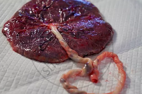
Placenta:
The placenta develops at the point of implantation. At 1st, the trophoblast cells absorb food and oxygen from the increasingly vascularised uterine lining. The allantois grows out from the embryo and fuses with the chorion to form the allanto-chorion which will develop into the placenta. The allantois gives rise to the umbilical cord which contains blood vessels connecting the foetus and placenta.
|
Placenta |
Function of Placenta:
- Nutrition: all the nutritive elements including glucose, fatty acid, amino acids, nucleotides, vitamins and minerals from the maternal blood pass into the foetus.
- Respiration: Oxygen passes from the maternal blood to the foetal blood through the placenta and CO2 pass in the reverse direction.
- Excretion: Excretory products diffuse into maternal blood and are again excreted by the mother.
- Storage: Stores glycogen, fat etc.
- Barrier: Allows only essential materials to pass into the foetal blood.
- Endocrine Function: Secretes hormones such as estrogen, progesterone, and human chorionic gonadotropin(hCG), hCS, and CRH.
Notes
Changes in embryo during pregnancy:
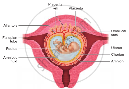
All these post-implantation steps lead to baby development. This takes 9 months for humans. Development is very slow and steady. First, the embryonic heart is formed, then the limbs, major organs, hair appearance, etc. At the end of 9 months, the foetus is fully developed. The limbs and external genital organs are well developed. The 1st movements of the foetus and the appearance of hair on the head are usually observed during the 5th month. By the end of 24 weeks the body is covered with fine hair, eyelids separate and eyelashes are formed. By the end of the 9th month of pregnancy, the foetus is fully developed and is ready for delivery.
|
Human foetus within the uterus |
Shaalaa.com | Human Reproduction Part 6
Related QuestionsVIEW ALL [43]
Match the columns I and II with reference to weeks of pregnancy and the development of a human embryo. Select the correct option from the choices given below:
| Column I | Column II | ||
| I. | 8 weeks | (P) | Limbs and external genital organs |
| II. | 12 weeks | (Q) | Limbs and digits develop. |
| III. | 20 weeks | (R) | Body hair develops. |
| IV. | 24 weeks | (S) | Eyelids separate. |
