Advertisements
Advertisements
प्रश्न
Distinguish between exonuclease and endonuclease
उत्तर
| Sr. No. | Exonuclease | Endonuclease |
|
1. |
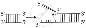
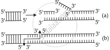
It is a type of restriction enzyme that removes the nucleotide from the 5’ or 3’ ends of the DNA molecule. |
It is a type of restriction enzyme that makes a cut within the DNA at a specific site to generate sticky ends. |
|
2. |
They separated DNA base pairs at their terminal ends.
|
They separate DNA at any location other than the terminal ends.
|
| 3. | They work on single strands of DNA or gaps in double-stranded DNA. | They cut one or both strands of double-stranded DNA. |
APPEARS IN
संबंधित प्रश्न
Explain with the help of a suitable example the naming of a restriction endonuclease.
Mention the difference in the mode of action of exonuclease and endonuclease.
Collect 5 examples of palindromic DNA sequences. Better try to create a palindromic sequence by following base-pair rules.
Give a reason why :
Single cloning site is preferred in a vector.
The total number of nucleotide sequences of DNA that code for a hormone is 1530. The proportion of different bases in the sequence is found to be Adenine = 34%, Guanine = 19%, Cytosine = 23%, Thymine = 19%.
Applying Chargaff’s rule, what conclusion can be drawn?
Restriction enzymes ______.
Molecular scissors, which cut DNA at specific site is ______.
DNA fragments separate according to size through?
Which of the given statements is correct in the context of visualizing DNA molecules separated by agarose gel electrophoresis?
'Restriction' in Restriction enzyme refers to ______.
While isolating DNA from bacteria, which of the following enzymes is not required?
The role of DNA ligase in the construction of a recombinant DNA molecule is ______.
What does H in’ ‘d’ and ‘III’ refer to in the enzyme Hind III?
Restriction enzymes should not have more than one site of action in the cloning site of a vector. Comment.
How does one visualise DNA on an agarose gel?
What is elution?
State the importance of elution in this process.
How are DNA fragments visualised once they are separated by gel electrophoresis?
